|
|
|
|
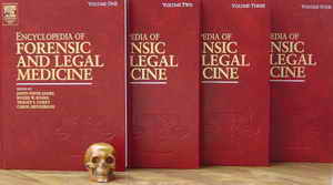
 Encyclopedia of Forensic and Legal Medicine (4 volumes), edited by Jason Payne-James, Roger Byard, Tracey Corey and Carol Henderson, Hard bound, 8.5 x 11
 Volume 1 - From Accreditation till Courts, Report writing - (xxxix + 495 pages)
Volume 2 From Crime-Scene Investigation and Examination till Human Rights Controls and Principles - (xxxix + 546 pages)
Volume 3 From Identification till Ritualistic Crime (xxxix + 564 pages)
Volume 4 From Road Traffic Accidents, Airbag related injuries and deaths till Yakuza plus author index (2 pages) plus subject index (87 pages) (xxxix + 479 pages)
 List of editors and editorial advisory board, two forewords (one by Bernard Knight giving a forensic perspective and another by Dame Elizabeth Butler-Sloss giving a legal perspective), preface, introduction, Guide to use of the encyclopedia, list of contributors and contents (of all four volumes) appear in each volume
 Price: $1095, £695.00, 995 [Introductory prices (till September 30th, 2005) - $740.00, £465.00, ¥105,039]
Visit the official site of this encyclopedia by clicking here.
Download contents of this encyclopedia by clicking here.
Buy from Amazon by clicking here.
 |
This is a landmark work, that the field of forensic medicine has seen in recent times. The board of editors decided to run some excerpts from this highly readable encyclopedia, so one could judge what a valuable addition to forensic literature it has been.
Several entries in this book deserve mention because they generally do not find place - or get just a passing mention - in ordinary pathology texts. The section "Animal attacks and injuries" has two entries - Fatal and Nonfatal written by AW Fegan-Earn of UK and Predation written by R. Rabinovich and Tzipi Kahana of Israel. In the latter entry, Rabinovich and Kahana describe injuries by insects and arachnids, marine animals, rodents, birds, small and large carnivores, large herbivores and reptiles. Among other things, we are told about the injuries by porcupines and birds, and how a forensic pathologist can identify them, how dogs can inflict blunt-force trauma and how they can cause injuries which could be confused with injuries associated with assault.
Here is what they have to say about injuries caused by rodents, birds and small carnivores..
ANIMAL ATTACKS AND INJURIES/PredationVolume 1, Pages 68-79
RodentsDeadly attacks by rodents are extremely rare; they are usually associated with small children of low socioeconomic background or debilitated persons. In these cases death, commonly caused by large species of rats, is due to blood loss resulting from multiple rat bites; subcutaneous bleeding around the wounds is the main indication that the injuries were inflicted while the victim was still alive. Autopsy findings often reveal signs of hypovolemic shock. Postmortem scavenging is common among wild and domestic rodents; they are well known to alter or destroy the indicators of the cause of death and preclude the visual identification of the victim. Rodents tend to gnaw on bone, to wear down on their incisors, leaving telltale sets of parallel striations on the osseous cortex. Postmortem rodent-caused injuries are usually wedged, paired, clean, small incisions without subcutaneous bleeding (Figure 4). Porcupines are known to collect and modify both dry and meaty bones. They leave a typical pattern of gnawed trails, thinning the bones in a fan-shaped pattern and creating "windows" in the shaft produced by heavy gnawing and scooping out material (Figure 5). Birds
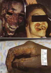
Serious injuries from birds resulting in death are very rare. The forensic literature mostly records postmortem stablike or puncture wounds caused by the hard bills of crows, owls, buzzards, or seagulls. Scavenging is a common feeding behavior of various species of birds, and remains are often completely defleshed in a matter of hours by these efficient foragers. Damage to bone occurs from the stripping and tearing action of the beaks and talons and small punctures and scratches on bone cortical surfaces are left. Finally, there have been instances where small birds have utilized the thoracic cavity of a skeletonized individual for nesting. Small Carnivores"Man's best friend" - the domestic dog - accounts for the majority of deaths caused by animal attacks. Attacks occur most often in the household domain, frequently against children and old adults. The majority of the lesions occur on the head, neck, and face, although they can be seen on the upper and lower extremities as well. The wounds are characterized by pairing of injuries resulting from the canine teeth, along linear parallel abrasions. Dogs can sometimes inflict blunt-force trauma by lifting their heads rapidly when excited, striking the victim in the throat area. Pets can sometimes paw their dead owners in an attempt to rouse them, inflicting groups of parallel abrasions that can be confused with injuries associated with assault.
Canids leave typical postmortem damage patterns over the bones, characterized by rounded punctures, peglike penetrating injuries, and shallow scratches. Severe damage caused by gnawing of the softer bony parts (proximal epiphyses) is frequently detected. Pack attacks by feral or wild dogs and wolves pose a greater danger of fatal injuries, similar to those produced by single animal attacks but often covering a larger area of the body. Not many reports have been related to deadly attacks of wolves on human; they are more often associated with attacks on flock animals. Overpopulation of foxes near or even in settled areas must be considered as dangerous. The scavenging behavior of packs of foxes can produce postmortem artifacts that can be easily misinterpreted as perimortem injuries. Large CarnivoresWild game attacks on humans occur mainly in rural areas, natural parks, zoos, and circuses. Large cats such as lions and tigers have been known to assault humans, although humans do not constitute their natural prey. The primary injuries sustained from these attacks are a series of parallel abrasions, multiple deep bite injuries, and lacerations with an abraded rim, consistent with the tapered claws of the animal. Punctured wounds similar in shape to those inflicted by smaller carnivores are often observed in the neck area of the victim. Shaking the victim by the neck is not uncommon, resulting in hypertension injuries. Furthermore, feeding on carcasses results in eventration (protrusion of the intestine through the abdominal wall), loss of tissue, and extensive bone injury.
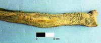
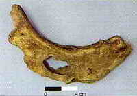
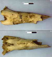
Attacks by hyena are uncommon, though they have been reported from Ethiopian villagers. Hyenas are known to plunder graves, dispersing the remains and leaving a puzzling scene for the unsuspecting investigator (Figure 6). Many carnivores crack bones with their teeth - the propensity of hyenids to feed on large diameter bones is legendary. Both species, spotted hyena (Crocuta crocuta) and striped hyena (Hyaena hyaena), tend to take chunks of their prey to their den, breaking and damaging the bones. Unique morphological features of hyena teeth and skull are associated with bone cracking. In addition to the morphological adaptations to lifting and carrying large and heavy loads, their ability to remove and destroy carcasses is noteworthy; scratches, furrows, puncture marks, and gnawed areas are typical of bone damage by hyena (Figures 7 and 8). |
||||
This chapter contains several other interesting photographs of injuries caused by all kinds of animals, with their descriptions and how pathologists could recognize them. Among other things, the authors have described instances where small birds have utilized the thoracic cavity of a skeletonized individual for nesting!
One section that is extremely informative is the one on Anthropology. It has 12 chapters under it (i) Overview (ii) Archaeology, excavation and Retrieval of Remains (iii) Taphonomy (iv) Stature estimation from skeleton (v) Bone pathology and ante mortem trauma (vi) Cremated bones (vii) Morphological age estimation (viii) Pediatric and Juvenile Anthropology (ix) Sex determination (x) Determination of Racial affinity (xi) Handedness and (xii) Role of DNA.
Truly representing the global spirit of the encyclopedia, this section is written by experts from several different nations. While Tzipi Kahana (Israel) writes the Overview, Archaeology is written by Haglund (USA) and Simmons ( UK), Stature Estimation by T Sjøvold (Sweden), Bone pathology and antemortem trauma by Sue M Black (UK), Cremated bones by P Holck (Norway), Morphological age estimation by Simpson (Australia) and Role of DNA by Ludes and Keyser-Tracqui (France). The reader is thus benefited from the combined experience of several researchers across the world, each a leader in his/her own field.
It would be worthwhile to excerpt a few paras from one of the chapters, so the reader can judge the quality of writing on his own. Here is what P. Holck of the University of Oslo, Norway has to say in his chapter on Cremated Bones.
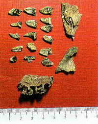
ANTHROPOLOGY/Cremated BonesVolume 1, Pages 113-119Bones Commonly Found after BurningWhen findings of cremated bones are examined, certain parts of the skeleton appear to be preserved more often than other structures. Bones from the neurocranium are commonly found as pieces of up to 10 cm in size, but bones from the facial skeleton are only occasionally found as such large fragments. This means that facial reconstructions of thermally decomposed bodies are unlikely to be possible. When an adult is exposed to temperatures of approximately 700°C, the face becomes a skeleton within 15 min. If the forehead is more or less intact, the frontal sinus should be examined and compared with X-rays of the presumed individual because the shape appears unique and thus this may be an important part of the identification work. Despite its thin and unprotected structure, the neurocranium is frequently found because of the insulating quality of the brain and cerebrospinal fluid. However, teeth, which are otherwise used to identify deceased persons, crack when they are exposed to heat. Enamel crowns are commonly lost while the roots remain intact; teeth of younger individuals usually resist heat better than those of elderly people. Thus, it is often not possible to use teeth of people exposed to fire for identification purposes (Figure 2).
The vertebral column is commonly well preserved: this may be related to the position of the body during burning. However, the sternum, ribs, clavicles, and scapulae are seldom seen in cremated material, probably because of their delicate shape and unprotected site in the body. Pieces of long bones can be found as fragments which are several centimeters long. Phalanges are seldom found in burnt forensic material, as opposed to in archeological finds, where these bones are seen relatively often: this may be explained by the local influence of cool open air as opposed to indoor fires. Also, the pelvis is seldom found, despite its protected site in the body, with the exception of pieces from the most solid parts. Human or Animal?In both forensic and archeological cases, there may be doubt as to the provenance of the bones: are the pieces human or not? Animals have much thicker, more compact bone than humans. Their trabecular units are a different size and shape, and this gives a heavier and more solid appearance than comparatively more porous and lighter human bones. The line between the spongy and the compact part of animal bone is also less distinctive than in human bones. Their outer/inner surface is often smoother and gives the examiner a feeling of unbreakable solidity. Some experts recommend microscopic examination to distinguish human from animal bones of uncertain origin, because the human Haversian canals are much wider than those of animal bones (Table 3).
In cremations, the examiner should pay more attention to macroscopic and morphologic-anatomic differences, even when limited, than to microscopic ones, because the bone shrinks and deforms, and can be very difficult to assess. Determination of SexThis is, of course, very important in all examinations. It should be based on common anatomical features, even if macro- and microscopic techniques have also been applied. It should be kept in mind that sexing cremated bones is very difficult and uncertain and requires more material than any other examination. It is common for more than 50% of cremated bone material to remain undetermined. This may be surprising to the police and others unfamiliar with such examinations, but it is always better to present a smaller number of certain determinations than a more impressive series based on suggestions. It is well known that the degree of sexual dimorphism varies between individuals and between different human races; nevertheless, the cremation process will sometimes emphasize sexually dimorphic criteria. Since larger and heavier parts of the skeleton, usually associated with men, are normally better preserved than smaller parts, some experts have suggested that sex criteria appear in cremated material as distinctly as in intact bones. Measurements and sexing based on mathematical or statistical methods should be used with the greatest care. As previously mentioned, the original shape of the bones shrinks and deforms in an unpredictable way. In fact, distinguishing features that are supposedly certain, such as the narrow male sciatic notch, may change into a wide, female-appearing structure after cremation in an incinerator. |
|||||||||||||||||||||
Order Encyclopedia of Forensic and Legal Medicine by Clicking here
Request a PDF file of this review by clicking here. (If your screen resolution can not be increased, or if printing this page is giving you problems like overlapping of graphics and/or tables etc, you can take a proper printout from a pdf file. You will need an Acrobat Reader though.)
Click here to read excerpts from this encyclopedia.
N.B. It is essential to read this journal - and especially this review as it contains several tables and high resolution graphics - under a screen resolution of 1600 x 1200 dpi or more. If the resolution is less than this, you may see broken or overlapping tables/graphics, graphics overlying text or other anomalies. It is strongly advised to switch over to this resolution to read this journal - and especially this review. These pages are viewed best in Netscape Navigator 4.7 and above.
-Anil Aggrawal


 [ Major links ]
[ Aims and Objectives ] [ FAQ ] [ Editorial Board ] [ Contributing Partners ] [ Sitemap ]
[ Paper/Thesis submission guidelines ] [ Editorials - Cumulative Index ] [ Be our sponsor! ]
[ Cumulative index of Book Reviews sorted by | Publishers | General Interest Books | Technical Books ] [ Animated Reviews ] [ Featured Reviews ]
[ Links ] [ Submit books/journals/software/multimedia for review ] [ journal CD ] [ History of the Journal ] [ Interviews ] [ Credits ]
[ Online Courses ] [ Awards ] [ Anil Aggrawal's Internet Journal of Book Reviews - Sister Publication ]
[ Cumulative reviews of Software/Multimedia | Books on CD/Audio tapes ] [ contact us ]
Books for review must be submitted at the following address.
Professor Anil Aggrawal (Editor-in-Chief)
Anil Aggrawal's Internet Journal of Forensic Medicine and Toxicology
S-299 Greater Kailash-1
New Delhi-110048
India
Click here to contact us.
This page has been constructed and maintained by Dr. Anil Aggrawal, Professor of Forensic Medicine, at the Maulana Azad Medical College, New Delhi-110002. You may want to give me the feedback to make this pages better. Please be kind enough to write your comments in the guestbook maintained above. These comments would help me make these pages better.
 IMPORTANT NOTE: ALL PAPERS APPEARING IN THIS ONLINE JOURNAL ARE COPYRIGHTED BY "ANIL AGGRAWAL'S INTERNET JOURNAL OF FORENSIC MEDICINE AND TOXICOLOGY" AND MAY NOT BE REPOSTED, REPRINTED OR OTHERWISE USED IN ANY MANNER WITHOUT THE WRITTEN PERMISSION OF THE WEBMASTER
Questions or suggestions ? Please use ICQ 19727771 or email to dr_anil@hotmail.com
Page Professor Anil Aggrawal via ICQ

|