|
|
|
|
 Endocrine Pathology- Differential Diagnosis And Molecular Advances, 1stEdition, Edited by Ricardo V. Lloyd. Hard Bound, 8.5" x 11".
Humana Press Inc., 999 Riverview Drive, Suite 208, Totowa, New Jersey 07512; Publication Date 2004. xii +421 pages, ISBN 1-58829-208-8; E-ISBN 1-59259-403-4 (acid-free Paper). List price $160 (discounts available on the Humana Press website)
 Official Site:Click here to visit
 |
Endocrine pathology forms an important part of surgical pathology reporting. Gone are the days when it was restricted to endocrine glands only. We now know that hormone-producing cells are widely distributed in the human body. These cells can produce a spectrum of lesion, which are great mimickers and often confuse a pathologist. This has been a battle of waterloo for every surgical pathologist. Endocrine pathology by Ricardo V Lloyd has been a great effort to solve these problems.
The book has 24 chapters and 12 pages of color plates. Each chapter is devoted to a specific organ. Each chapter discusses normal histology followed by pathological lesions. This is followed by an exclusive chapter on recent developments in molecular biology wherever applicable. The first chapter: "Differential diagnosis and molecular advances" discusses all the basic methods related to research and diagnosis. The diagrammatic representations are very elegant and crisp. The illustrations are adequate and very appropriate making understanding easy.
 |
Both the techniques and diagnostic utility of molecular biology have been dealt with in great detail. It is up-to-date and well discussed. The following paragraph from the book unambiguously summarizes the molecular approach of this book:
"The problem of cellular heterogeneity has been a major barrier to the molecular genetic analysis of normal versus diseased tissue. Thus tissue micro-dissection represents one of the most promising techniques in molecular pathology offering a link between morphology and molecular genetic analysis" (Page 18, last para).
Lloyd et al has tried to weave a link between morphology and molecular genetics. It is extremely admirable the way they have done it.
After going through the book one is left with no option but to agree with Lloyd's statement that "Today's state-of-the-art concept will soon become obsolete with new developments in cell and molecular biology. However the basic time-tested concepts in diagnostic endocrine pathology should remain relevant and continue to serve as the spring board from which the most important new discoveries in this field will be launched over the next few years".
 |
However, one feels that "this basic time-tested concept" of morphological pathology would have been served better if better quality microphotographs particularly more of colored photographs were illustrated. Since, histopathology is heavily based on photogenic memory and most surgical pathologists are used to making histologic diagnosis of unusual lesions by photo-matching, good and clear photographs are extreme necessity in any pathology book (An instance of this occurs in fig. 12 on p 177, where it is difficult to see the microfollicular structure). Most of the microphotographs do not give any magnification. In its place, a bar has been used. As a pathologist, one is more familiar with magnifications. Concept of bar is good for gross photographs which are surprisingly scanty.
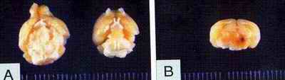
The book is illustrated with some very good color photographs. Look for instance at the gross photograph (above left) which shows pituitary adenoma (Color plate 3, Figure 9). A reader would greatly appreciate the gross of different endocrine tumors, which at times can be differentiated, from non-endocrine tumors by color. However black-and-white photographs are hardly of any use in immunohistology, least of all in double staining (One such instance occurs on page 6, Fig. 8). Though color plates have been provided, but flipping back and forth to match the photographs with the text may at times become boring and irritating more so when the color plates do not carry any legend.
 |
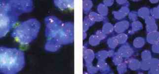 |
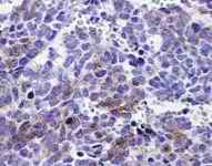
On right we can see some more illustrative photographs from this book. The one on the extreme left shows synaptophysin in pineoblastoma and the next two photographs show FISH in parathyroid carcinoma and adenoma respectively. In my humble opinion, it would have been better to put the color photographs along with their text. These changes may increase the price a bit, but will be far more useful and saleable.
However, the book is remarkable for its up-to-date knowledge and details. It has also dealt with contemporary problems such as obesity and anorexia nervosa in the chapter "Appetite regulation, obesity and anorexia nervosa". Similarly, pituitary and parathyroid lesions are also discussed well. Parathyroid adenoma has been written very well. However, a common problem of distinguishing a parathyroid adenoma from hyperplasia needs to have been touched in little more detail rather than stating - "the main differential diagnosis of parathyroid adenoma and nodular hyperplasia is that it shows multiple gland involvement". Thyroiditis has been split into too many subtype e.g. postpartum thyroiditis and silent thyroiditis. Though these hair-splitting variants are being reported but a line of caution should have been added that these are basically variants of other types of thyroiditis. Two important types of thyroiditis - lymphocytic and Hashimoto's thyroiditis are possibly two poles of the same entity.
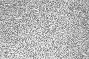 |
Table number 5, chapter 9, page 169 very clearly illustrates the differences between hyperplasia, follicular adenoma, follicular carcinoma and follicular variant of papillary carcinoma. A simple morphological appearance of scalloping, thin colloid and pseudopapillary formation may also differentiate hyperplasia from the rest. Paraganglioma has been written extremely well.
The figure on the left shows spindle cell variant of paraganglioma. Neuroendocrine lesions of multiple organs such as lung, breast, skin and urogenital tract have been elaborately discussed. However, bone tumors (Primitive Neuroendocrine tumor, PNET) do not seem to have caught Dr. Ricardo V Lloyd's fancy in this otherwise very exhaustive book. One also wonders why parasitic and fungal diseases have been denied an entry in this book which are increasingly becoming important due to AIDS. Readers will also feel handicapped searching many entities from the index.
Overall, it is a well-written, up-to-date and fairly exhaustive book. A book which any surgical pathology section must have.
|
Dr. A.K.Mandal is a graduate of Maulana Azad Medical College, New Delhi. He did his post graduation in Pathology from All India Institute of Medical Sciences, New Delhi. Later, he went on to do his PhD from Fukushima Medical University, Japan. His further training included a Commonwealth Fellowship at the Institute of Genetics, University of Wales, Cardiff, U.K. He was WHO fellow at the University of Texas, Huston. He joined Maulana Azad Medical College as a faculty and has been involved in Surgical Pathology since last twenty-five years. Dr Mandal is one of the most respected and distinguished oncopathologists on the Asian continent. He is currently heading the Surgical Pathology unit at Maulana Azad Medical College, New Delhi. He has been a vice-president of National Board of Examinations and is on the editorial board of a number of journals. A superb teacher and an exemplary pathologist, Dr. Mandal has maintained an intense devotion to research. |
-Anil Aggrawal
[ Aims and Objectives ] [ FAQ ] [ Editorial Board ] [ Contributing Partners ] [ Sitemap ]
[ Paper/Thesis submission guidelines ] [ Editorials - Cumulative Index ] [ Discussion ] [ Chat room ] [ Be our sponsor! ]
[ Cumulative index of Book Reviews sorted by | Publishers |
General Interest Books |
Technical Books ] [ Animated Reviews ] [ Featured Reviews ] [ E-books ]
[ Reviews with Quizzes ] [ Links ] [ Submit books/journals/software/multimedia for review ] [ journal CD ] [ History of the Journal ] [ Interviews ] [ Credits ]
[ Cumulative index of | Theses/dissertations | [ Online Courses ] [ Awards ] [ Anil Aggrawal's Internet Journal of Book Reviews - Sister Publication ]
[ Cumulative reviews of Software/Multimedia | Books on CD/Audio tapes ] | Calenders | Models ] [ contact us ]
[ Undergraduate section | Postgraduate section ] [ Forensic gadgets/toys/other tidbits ]
Questions or
suggestions ? Please use ICQ 19727771
or email to
dr_anil@hotmail.com
Page Professor Anil Aggrawal via
ICQ
Order Humana Press Books by clicking here.
 Request a PDF file of this review by clicking here. (If your screen resolution can not be increased, or if printing this page is giving you problems like overlapping of graphics and/or tables etc, you can take a proper printout from a pdf file. You will need an Acrobat Reader though.)
N.B. It is essential to read this journal - and especially this review as it contains several tables and high resolution graphics - under a screen resolution of 1600 x 1200 dpi or more. If the resolution is less than this, you may see broken or overlapping tables/graphics, graphics overlying text or other anomalies. It is strongly advised to switch over to this resolution to read this journal - and especially this review. These pages are viewed best in Netscape Navigator 4.7 and above.


 [ Major links ]
Books for review must be submitted at the following address.
Professor Anil Aggrawal (Editor-in-Chief)
[ Major links ]
Books for review must be submitted at the following address.
Professor Anil Aggrawal (Editor-in-Chief)
Anil Aggrawal's Internet Journal of Forensic Medicine and Toxicology
S-299 Greater Kailash-1
New Delhi-110048
India
Click here to contact us.
This page has been constructed and maintained by Dr. Anil Aggrawal, Professor of Forensic Medicine, at the Maulana Azad Medical College, New Delhi-110002. You may want to give me the feedback to make this pages better. Please be kind enough to write your comments in the guestbook maintained above. These comments would help me make these pages better.
 IMPORTANT NOTE: ALL PAPERS APPEARING IN THIS ONLINE JOURNAL ARE COPYRIGHTED BY "ANIL AGGRAWAL'S INTERNET JOURNAL OF FORENSIC MEDICINE AND TOXICOLOGY" AND MAY NOT BE REPOSTED, REPRINTED OR OTHERWISE USED IN ANY MANNER WITHOUT THE WRITTEN PERMISSION OF THE WEBMASTER

home
> Volume 5, Number 2, July - December 2004
> Reviews
> Technical Books
> Page 2: Endocrine Pathology- Differential Diagnosis And Molecular Advances
> page 2a: Endocrine Pathology (Review by Dr. A.K.Mandal of India) (you are here)
Navigation ribbon
![]()