Paper 1: Orbital Index of Adult Malawians by Igbigbi and Ebite: Anil Aggrawal's Internet Journal of Forensic Medicine: Vol. 11, No. 1 (January - June 2010)
Received: June 26, 2008
Revised paper received: June 20, 2009
Accepted: June 28, 2009
Ref:
Igbigbi, P.S., Ebite, L.E. Orbital Index of Adult Malawians.
Anil Aggrawal's Internet Journal of Forensic Medicine and Toxicology [serial online], 2010; Vol. 11, No. 1 (January - June 2010): [about 21 p]. Available from: . Published : January 1, 2010, (Accessed:
Email the corresponding author P.S.Igbigbi by clicking here
Patrick S. Igbigbi
Lilian Ebele Ebite
Orbital Index of Adult Malawians
by Professor Patrick S. Igbigbi, MB.BS, M.Sc, M.D
Professor of Neuroanatomy and Physical Anthropology
Delta State University Abraka Delta State Nigeria
P.M.B 1 Abraka Delta State Nigeria
Formerly Dean College of Medicine
University of Malawi
Blantyre Malawi
and
Dr Lilian Ebele Ebite MB.BS, MSc
Lecturer, Department of Anatomy,
Delta State University
EMAIL: lilyzoks@yahoo.com
Phone no: +2348056614585
E-mail for correspondence: pigbigbi@yahoo.com
Abstract
The orbital height, width, and index of 136 anteroposterior skull radiographs of adult Malawians aged between 18-73 yrs were analysed retrospectively. Results showed that mean orbital width was higher in males (44.57±2.24 mm) than females (42.15±2.20 mm) but the females had a higher mean orbital index (96.03±3.34 mm). The mean orbital width was higher in females than males within the age group 18-27 years and 38-47 yrs but the reverse was the case in the other adult age groups---28-37, 48-57, 58-67, 68-77 years. The mean orbital height showed a steady rise in males with increasing age to a maximum of 45.00 mm at 58-67 years followed by a slight decline at 68-77 years age group; suggesting adult variations of fused bone size probably due to bone remodelling with age. The results show that adult Malawians belong to the megaseme group just like the Chinese and Polynesians.
Keywords
Orbital Index, Orbital Height, Orbital Width, Malawians, Radiographs
Introduction
The orbital cavities are situated on either side of the sagittal plane of the skull between the cranium and the skeleton of the face.1 Each orbital cavity is essentially intended as a socket for the eye-ball and also contains associated muscles, nerves, vessels and in essence lodges the visual apparatus.2 This anatomical region is therefore of clinical and surgical interest in ophthalmology, oral and maxillofacial surgery and neurosurgery.1
The orbital margin is quadrilateral in shape with rounded corners and usually has a spiral form. The inferior margin is continuous with the anterior lacrimal crest , while the superior is continued down into the posterior lacrimal crest and the fossa thus formed lies in the orbital margin.
Each orbital cavity measures about 40mm with the width usually greater than the height, the relation between the two is given by the orbital index, which varies in the different races of mankind.1
The Orbital Index =
Height of orbit
X 100
Width of orbit
Taking the orbital index as the standard, three classes of orbits are recognized.
• Megaseme (large) - The orbital index is 89 or over. This type is characteristics of the yellow races, except the Esquimaux (Eskimos) where the orbital opening is round.3,4
• Mesoseme (intermediate) - The orbital index range between 89 and 83. This type is found in the white races (European 87, English 88.4).5
• Microseme (small) - Orbital index 83 or less. This type is characteristics of the black races where the orbital opening is rectangular. 6
This index which determines the shape of the face differs in different population groups. This means that the orbits with larger widths than height will have smaller orbital indices while those with larger orbital indices will have narrow faces. This index varies with race, regions within the same race and periods in evolution. For example, the orbital index seen in modern man of the Kanto region and Kinki regions of Japan were of the microseme range (79.26-80.33); In Peking province of China, studies showed orbital index of microseme category(80.68), Fushun region - mesoseme category (83.57) while Hokien region showed megaseme orbital index (90.39), an indication of regional variability of the index in China.
7,8,9,10,11
Furthermore, measurements of the Minatogawa prehistoric men of Japan showed orbital indices of 65.2-66.7, and the Neolithic men 76.39-75.11. Apparently, the orbital width reduced in the process of evolution while the height increased. This means a rearrangement towards a longer slimmer face in the modern Japanese man.7
The knowledge of this index is therefore important in various aspects such as in interpretation of fossil records, skull classification in forensic medicine and in exploring the trends in evolutionary and ethnic differences. Furthermore documented ranges of this index in different nationalistic groups will assist in skull identification.12,13
This study is therefore aimed at filling this anthropometric gap among adult Malawians.
Materials and Methods
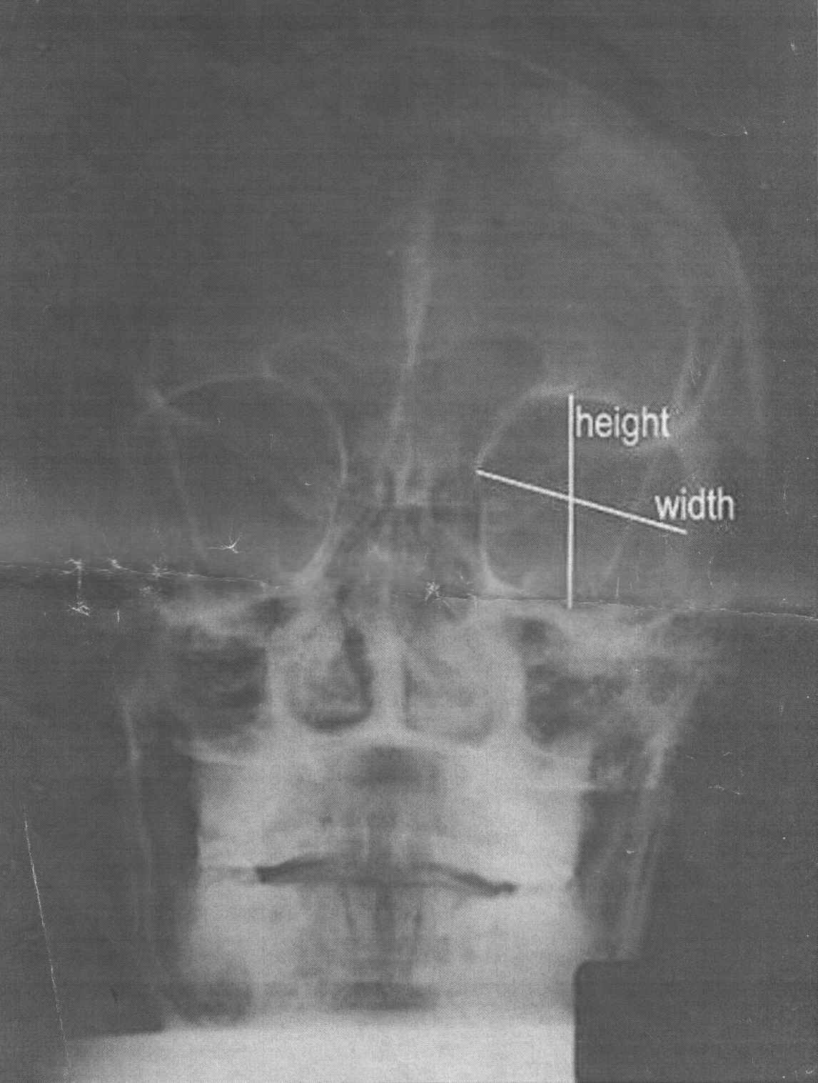
We analysed 136 anteroposterior radiographs (Waters' view) of adult Malawians aged between 18 -73 years. Adult skull x-rays were used because it was assumed that all the skull bones have fused unlike in young skulls which are still growing.
Figure 1. A typical X-ray used in this study. Please note how height and width were taken from X-rays.
(Click picture to enlarge)
Radiographs were collected from Queen Elizabeth central hospital and Mulamulo Adventist hospital both of which were referral hospitals dealing with orthopaedic patients in Malawi.
Permission to access the x-rays was given by the directors and heads of radiology department of the various hospitals, and only x-rays with occipito-mental views were used (Figure-1).
A total of 187 X-rays were obtained from both hospitals and of this only 136 (66 females and 70 males) were correctly visible and hence used. We then classified each radiograph according to the age and gender of the individual, as provided on the radiograph jackets. A 30mm ruler was used to measure the width and height of the orbits with the skull X-ray on the X-ray viewer and the orbital index was calculated using the formula indicated previously.
Results
The results are summarized in three tables. Table 1 Shows the orbital height, widths and indices for male and female adult Malawians. The indices for both sexes was found to be higher than 89.00. Total mean orbital height was 40.26 in females and 42.07 in males. Total mean orbital width was 42.15 in females and 44.57 in males. Total orbital index varied more in males than females with the mean total orbital index in females 96.03 in females while in males it was 94.35.
Table 1: Summary of Orbital Parameters (Width, Height And Index) in the Present Study
Sex
Number
Total mean height (mm)
Total mean width (mm)
Total orbital index
Male
70
42.07(2.96)
44.57(2.24)
94.35 + 5.56
Female
66
40.26(1.92)
42.15(2.20)
96.03 +3.34
Total
136
41.17(2.44)
43.36(2.22)
95.19+ 4.56
What is already known on this topic
Taking the orbital index as the standard, three classes of orbits are recognised.
. Megaseme (large) - The orbital index is 89 or over. This type is characteristics of the yellow races, except the Esquimaux (Eskimos) where the orbital opening is round.
. Mesoseme (intermediate) - The orbital index range between 89 and 83. This type is found in the white races (European 87, English 88.4)
. Microseme (small) - orbital index 83 or less. This type is characteristics of the black races where the orbital opening is rectangular.
What This study adds
This study showed that mean orbital width was higher in males (44.57±2.24) than females (42.15±2.20) but the females had a higher mean orbital index (96.03±3.34). The mean orbital width was higher in females than males within the age group 18-27 years and 38-47 yrs but the reverse was the case in the other adult age groups - -28-37, 48-57, 58-67, 68-77 years. The mean orbital height showed a steady rise in males with increasing age to a maximum of 45.00 cm at 58-67 years followed by a slight decline at 68-77 years age group, Suggesting adult variations of fused bone size probably due to bone remodelling with age. The result show that adult Malawians belong to the megaseme group just like the Chinese and Polynesians.
This study present for the first time the orbital parameters in adult Malawians thus providing a useful baseline and an anthropometric data that will be of clinical and surgical interest in ophthalmology and oral and maxillofacial surgery and indeed neurosurgery.
Suggestions for further development
The orbital parameters obtained from roentgenographs has been shown to be slightly different from those obtained from direct measurement of human skulls and this difference could be attributed to magnification factor of x-ray machines hence in obtaining a roentgenograph, angle of emission of radiation, distance from source of radiation and positioning of the patient must all be standardised to give valid reproducible results.
Tables 2A & B Show the spread of the mean orbital heights, width and indices with different male adult age groups. The highest mean orbital heights occurred at 48-77 age group (43.25mm-45mm). The widest mean orbital width occurred at 48-77 years (45.00mm-47.33mm), while the smallest orbital dimensions of height (38.45mm) and width (40.85mm ) occurred at age group 18-27 years and the largest orbital dimensions of height (44.00mm and width 47.33mm occurred at age group 68-77 years. Width -height difference, arranged in ascending order with mean orbital index has been shown in table 2B.
Table 2A: Age Group
Distribution of Orbital Parameters in Males
Number in each age group
Age group
Mean orbital height (mm)
Mean orbital width(mm)
Difference height & width
(mm)
Mean orbital index
20
18-27
38.45(6.62)
40.85(2.85)
2.40
94.07(15.4)
25
28-37
40.50(4.18)
43.85(4.08)
3.35
92.36(6.22)
17
38-47
41.19(3.06)
43.41(2.99)
2.22
94.88(5.88)
4
48-57
43.25(1.26)
45.00(1.41)
1.75
96.11(1.08)
1
58-67
45.00(0.00)
47.00(0.00)
2.00
95.74(0.00)
3
68-77
44.00(2.65)
47.33(2.08)
3.33
92.04(4.76)
Table 2B : Age Group
Distribution of Width Height Difference and Orbital Index in Males
Difference between height and
width( in ascending order)
Mean orbital index
Age group
1.75
96.11
48-57
2.00
95.74
58-67
2.22
94.88
38-47
2.40
94.07
18-57
3.33
92.96
68-77
3.35
92.36
28-37
Tables 3A&B show the spread of mean orbital height with orbital index and width /height difference with age groups. The highest mean orbital height occurred at age group 38-47 years (39.93-44.47mm) while the highest mean orbital width occurred at age group 58-67 years (44.60mm-47.4mm).
The smallest orbital dimensions of 41.00mm height, 38.00mm width occurred at 68-77 years, while the highest orbital dimensions of 42.20mm height, 43.67mm width occurred at 38- 47 years. Similarly the width -height difference, arranged in ascending order compared with their corresponding orbital index as shown in table 3B.
Table 3A
: Age Group Distribution of Orbital Parameters in Females
Number in each group
Age group in years
Mean orbital height
Mean orbital width(mm)
Difference btw height & width
Mean orbital index
14
18-27
39.86(2.17)
41.79(2.12)
1.93
95.58(6.52)
27
28-37
39.52(3.59)
41.70(3.21)
2.18
94.77(3.65)
15
38-47
42.20(2.27)
43.67(2.55)
1.47
96.63(4.47)
7
48-57
41.00(3.51)
41.71(3.90)
0.71
98.30(3.35)
2
58-67
38.00(0.00)
46.00(1.40)
8.00
82.61(2.25)
1
68-77
41.00(0.00)
38.00(0.00)
-3.00
107.89(0.00)
Table 3B : Age Group
Distribution of Width Height Difference And Orbital Index in Females
Difference btw height and width(
in ascending order)
Mean orbital index
Age groups
-3.00
107.89
68-77
0.71
98.30
48-57
1.47
96.63
38-47
1.93
95.58
18-27
2.18
94.77
28-37
8.00
82.61
58-67
Discussion
From the study, the mean orbital index of adult Malawians was found to be 95.15±4.56 (90.63-99.75). This value places the orbital index of adult Malawians in megaseme category. Other races with orbital height as close to the width in magnitude as possible (square orbits) include the Chinese as indeed have been shown to be the characteristics of the yellow races.1 However a previous study has demonstrated that the black races have microseme orbital index.1
Furthermore the orbital indices of females were generally higher than males showing that female orbits were closer to having an orbital index of 100 (square shaped) than males for the same age group. It is also noteworthy that the three orbital roentogenographs of females in age group 58-71 showed widely varying values of orbital height and weight. The only case in which orbital height had greater magnitude than orbital width showing that although adult Malawians orbits are in megaseme category, adult female Malawian orbits are more megaseme than the male. The difference between the orbital indices of adult females and males with the age groups 18-57 years was on the average 1.965 + 0.35 showing that within the same age group female orbital indices were higher than males by a factor of almost 2.
Another noticeable thing was the variation in orbital dimensions with age. This suggest either a genetically determined continuously variable like height or may be due to continuous bone resorption and remodelling which according to Parfitt14,15
occurred at cortical bone surface every 2-5 years while bone turnover for the whole skeleton is about 10% per years.
Mean orbital height of male Malawian showed a more or less steady increase with age; A similar increase was noticed with mean orbital width in males with the largest orbit occurring at age 68-77 years. This reflects an age dependent remodelling of bone, this was in agreement with Uberlaker,16 Stout17 and Castanet et al.18 This degree of remodelling was in general an indication of age so that the number of osteons and osteon fragment had been used in attempts to estimate the age of skeletal materials at death.16 This remodelling apparently drives toward a longer face with age in male Malawians.
Conversely female Malawians in this study showed mean orbital heights and widths which do not follow a more or less clear pattern like those of males. The pattern seem more unusual at older ages of 58-77 with width -height difference varying more widely then. In fact we found a single female roentgenograph with orbital height greater than orbital width this variations was probably due to the effects of oestrogens and androgens on bone resorption and remodelling of bone. At menopause 45-56 years there was withdrawal of oestrogen leading to increased bone resorption at this period. Expectedly at this age many of the adult female could be on calcium tablets so as to minimally reduce the effects of oestrogen withdrawal.
The orbital width /height difference bears a marked relationship to mean orbital index seen clearly when arranged in ascending order of "width /height difference" The mean orbital index in an age group bears an inverse relationship to the width /height difference for that age group. We are reporting this relationship probably for the first time.
A look at (appendix 1-using a difference of zero (0) as equivalent to mean orbital index of 100 and interval of 2.34. We derived a table which correctly relates the values obtained in this study. This value of 2.34 was obtained by dividing the difference between the highest and lowest mean orbital index by the difference between the highest and lowest width /length difference.
A look at appendix 1 shows that when the range of width-height difference is between 0.0-1.0, the equivalent range of mean orbital index is 100.0-97.66 (interval of 2.34). This value of 2.34 was obtained by dividing the difference between the highest and lowest mean orbital index by the difference between the highest and lowest width /length difference.
Appendix 1
Width-Height Difference
and Mean Orbital Index
Range of
width-height difference
Equivalent
range of mean orbital index
(-2.1)- (-3.0)
104.69-107.02
(-1.1)- (-2.0)
102.35-104.62
(-0.1)- (-1.0)
100.10-102.34
0.0- 1.0
100.0-97.66
1.1-2.0
97.65-93.32
2.1- 3.0
95.31-92.98
3.1-4.0
92.97-90.64
4.1-5.0
90.63-88.30
5.1-6.0
88.29-85.96
6.1-7.0
85.95-83.62
7.1- 8.0
83.61-81.28
The orbital parameters obtained from roentgenographs has been shown to be slightly different from those obtained from direct measurement of human skulls and this difference could be attributed to magnification factor of X-ray machines hence in obtaining a roentgenograph, angle of emission of radiation, distance from source of radiation and positioning of the patient must all be standardised to give valid reproducible results.19
This study presents for the first time the orbital parameters in adult Malawians thus providing a useful baseline and an anthropometric data that will be of clinical and surgical interest in ophthalmology, oral and maxillofacial surgery and indeed neurosurgery in this part of the world.
N.B. It is essential to read this journal - and especially this paper as it contains several tables and high resolution graphics - under a screen resolution of 1600 x 1200 dpi or more, and preferably on a 17" or bigger monitor. If the resolution is less than this, you may see broken or overlapping tables/graphics, graphics overlying text or other anomalies. It is strongly advised to switch over to this resolution to read this journal - and especially this paper. These pages are viewed best in Netscape Navigator 4.7 and above.
This page has been constructed and maintained by Dr. Anil Aggrawal, Professor of Forensic Medicine, at the Maulana Azad Medical College, New Delhi-110002. You may want to give me the feedback to make this pages better. Please be kind enough to write your comments in the guestbook maintained above. These comments would help me make these pages better.
IMPORTANT NOTE: ALL PAPERS APPEARING IN THIS ONLINE JOURNAL ARE COPYRIGHTED BY "ANIL AGGRAWAL'S INTERNET JOURNAL OF FORENSIC MEDICINE AND TOXICOLOGY" AND MAY NOT BE REPOSTED, REPRINTED OR OTHERWISE USED IN ANY MANNER WITHOUT THE WRITTEN PERMISSION OF THE WEBMASTER
 Igbigbi, P.S., Ebite, L.E. Orbital Index of Adult Malawians.
Anil Aggrawal's Internet Journal of Forensic Medicine and Toxicology [serial online], 2010; Vol. 11, No. 1 (January - June 2010): [about 21 p]. Available from:
. Published : January 1, 2010, (Accessed:
Igbigbi, P.S., Ebite, L.E. Orbital Index of Adult Malawians.
Anil Aggrawal's Internet Journal of Forensic Medicine and Toxicology [serial online], 2010; Vol. 11, No. 1 (January - June 2010): [about 21 p]. Available from:
. Published : January 1, 2010, (Accessed:
 Email the corresponding author P.S.Igbigbi by


 This page has been constructed and maintained by Dr. Anil Aggrawal, Professor of Forensic Medicine, at the Maulana Azad Medical College, New Delhi-110002. You may want to give me the feedback to make this pages better. Please be kind enough to write your comments in the guestbook maintained above. These comments would help me make these pages better.
 IMPORTANT NOTE: ALL PAPERS APPEARING IN THIS ONLINE JOURNAL ARE COPYRIGHTED BY "ANIL AGGRAWAL'S INTERNET JOURNAL OF FORENSIC MEDICINE AND TOXICOLOGY" AND MAY NOT BE REPOSTED, REPRINTED OR OTHERWISE USED IN ANY MANNER WITHOUT THE WRITTEN PERMISSION OF THE WEBMASTER

