Poster Session: POSTER 4: Metastasis in heart from a primary carcinoma of hypopharynx -A rare event by B.D. Gupta and colleagues :Anil Aggrawal's Internet Journal of Forensic Medicine, Vol.10, No. 2, July - December 2009
Received: January 25, 2009
Accepted: March 29, 2009
Ref: Gupta B.D., Vaghela, P.C., Singh, O.G. and Mehta, R. Metastasis in heart from a primary carcinoma of hypopharynx -A rare event. Anil Aggrawal's Internet Journal of Forensic Medicine and Toxicology, 2009; Vol. 10, No. 2 (July - December 2009):
; Published: July 1, 2009, (Accessed:
Gupta B.D., Vaghela, P.C., Singh, O.G. and Mehta, R. Metastasis in heart from a primary carcinoma of hypopharynx -A rare event. Anil Aggrawal's Internet Journal of Forensic Medicine and Toxicology, 2009; Vol. 10, No. 2 (July - December 2009):
; Published: July 1, 2009, (Accessed:
Email Dr. B.D.Gupta (the corresponding author) by clicking here

|
Dr. B.D. Gupta (Click to enlarge)
Poster Session: Poster 4
Metastasis in heart from a primary carcinoma of hypopharynx -A rare event
|
B. D. Gupta*, P. C. Vaghela**, O. G. Singh**, Rahul Mehta***
* Prof & Head, **Tutor, ***PG Student
Forensic Medicine Department
M.P.Shah Medical College, Jamnagar
India
|
Case Report
|
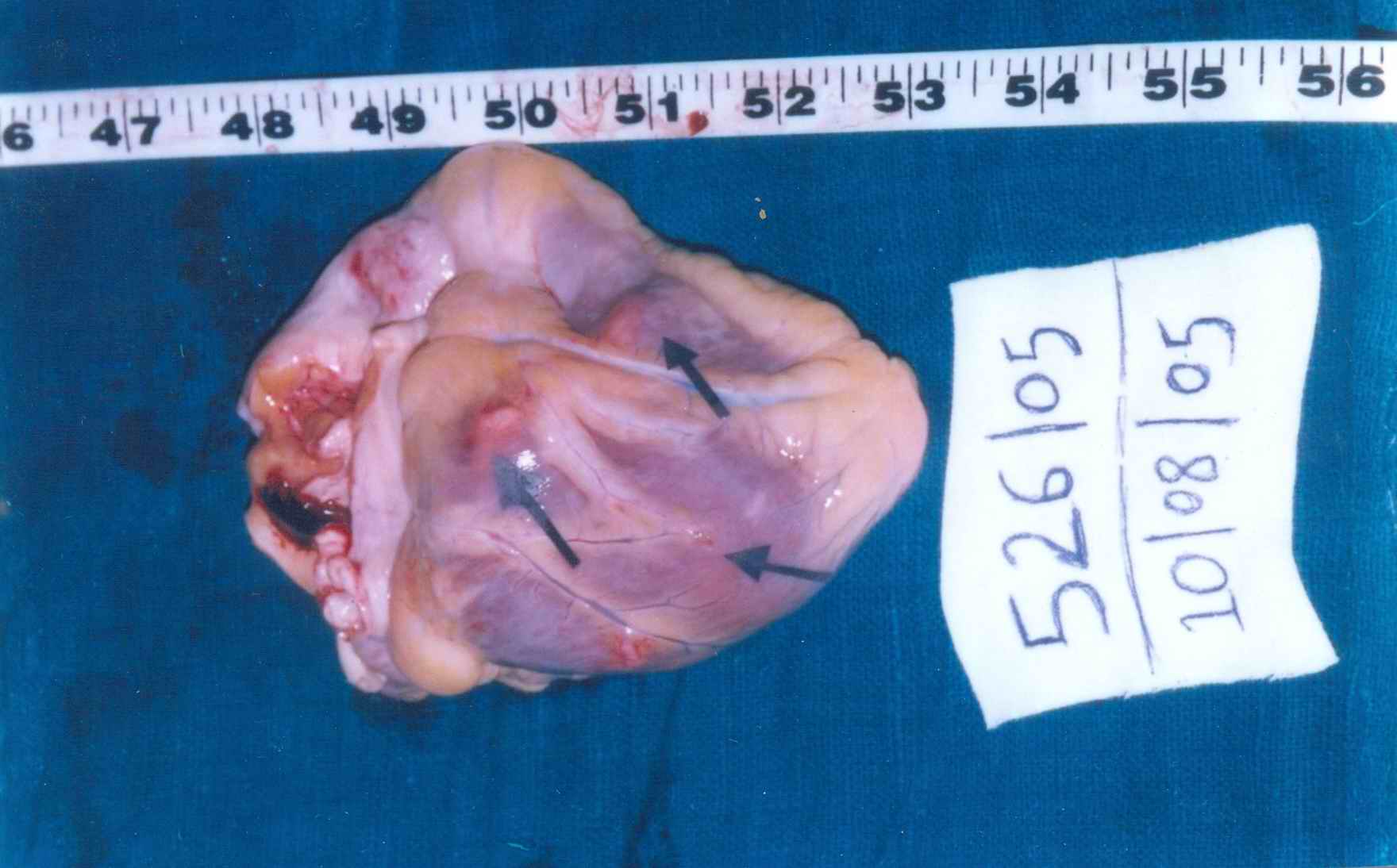
Picture 1: Heart shows multiple nodules of metastasis (Please see arrows)
|
|
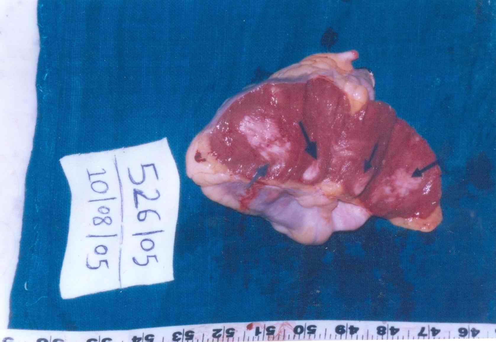
Cut surface of heart shows multiple nodules of metastasis. (Please see arrows)
|
|
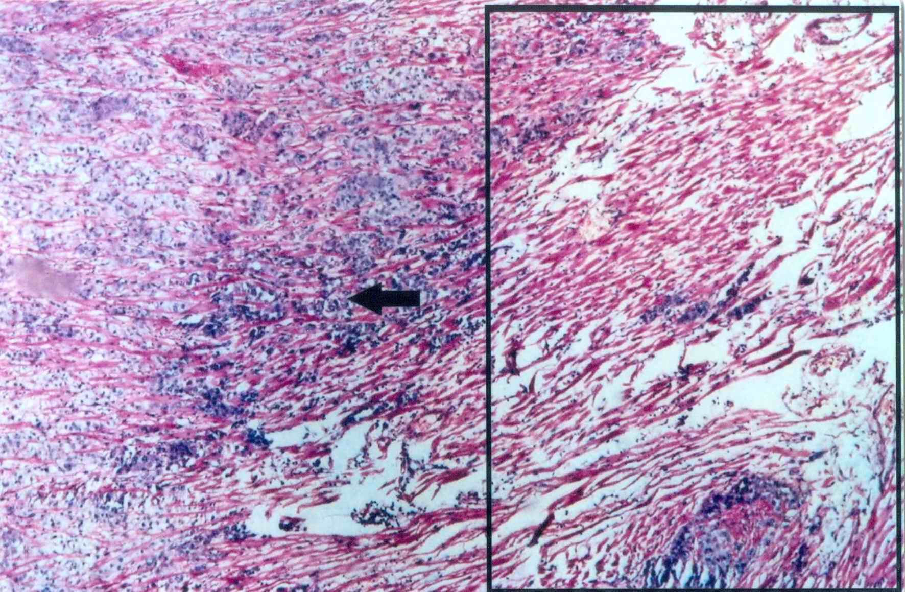
Picture 3: Heart muscles infiltrated with tumour cells. (x 40) Rectangular area shows heart tissue. Arrow shows infiltration of tumour cells.
|
|
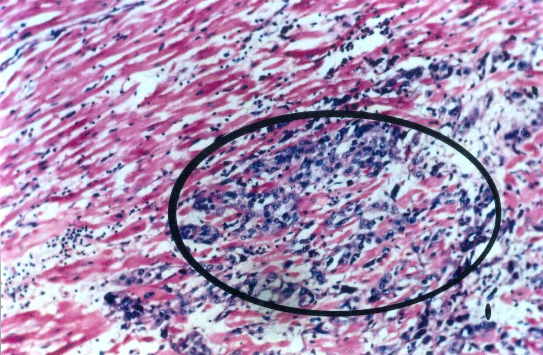
Picture 4: Heart muscles infiltrated with tumour cells. (x 100). Oval area shows infiltration of tumour cells. Region outside the oval area shows heart tissue.
|
|
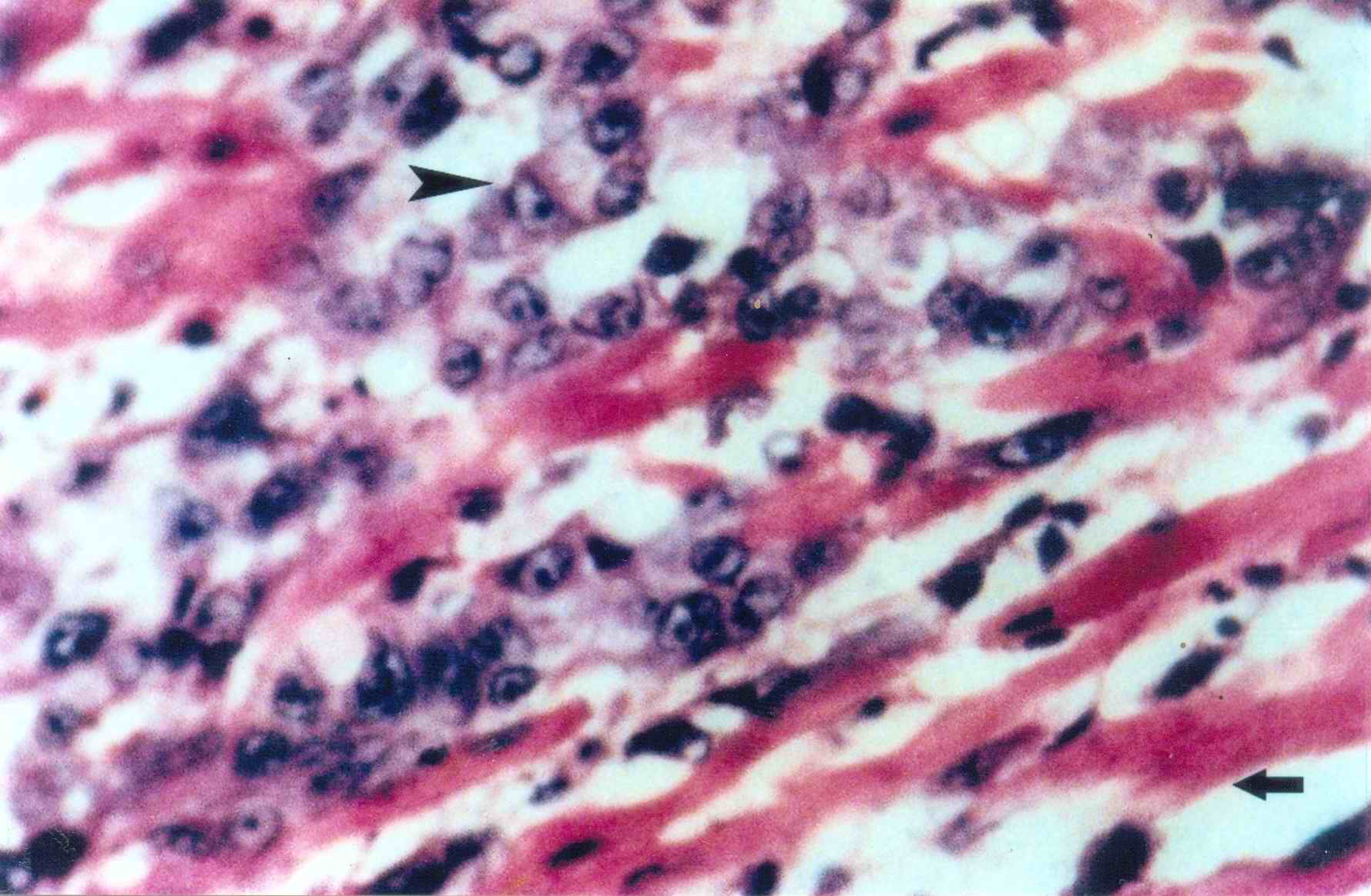
Picture 5: Heart muscles and tumour cells. (x 400). Arrow top left shows infiltration of tumour cells. Arrow bottom right shows heart tissue.
|
|
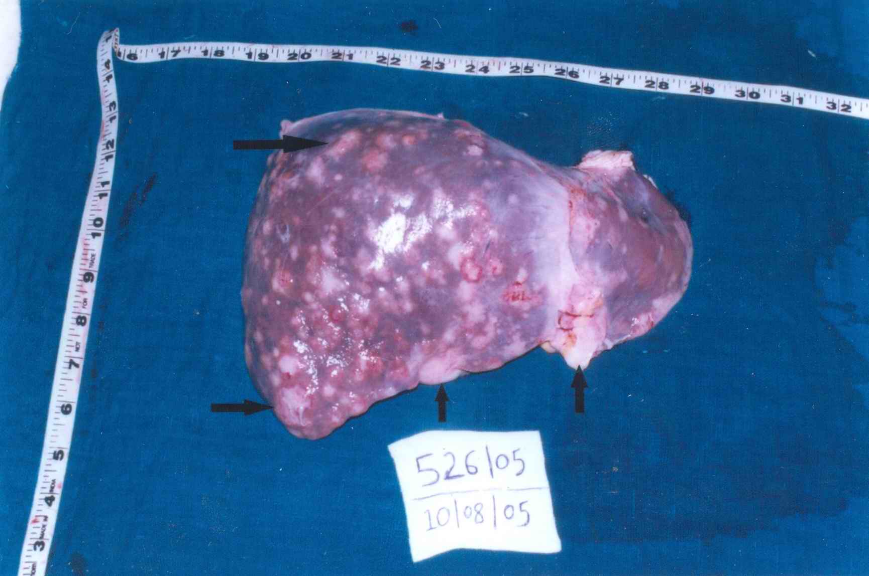
Picture 6: Liver (gross) shows multiple nodules of metastasis. (Please see arrows).
|
|
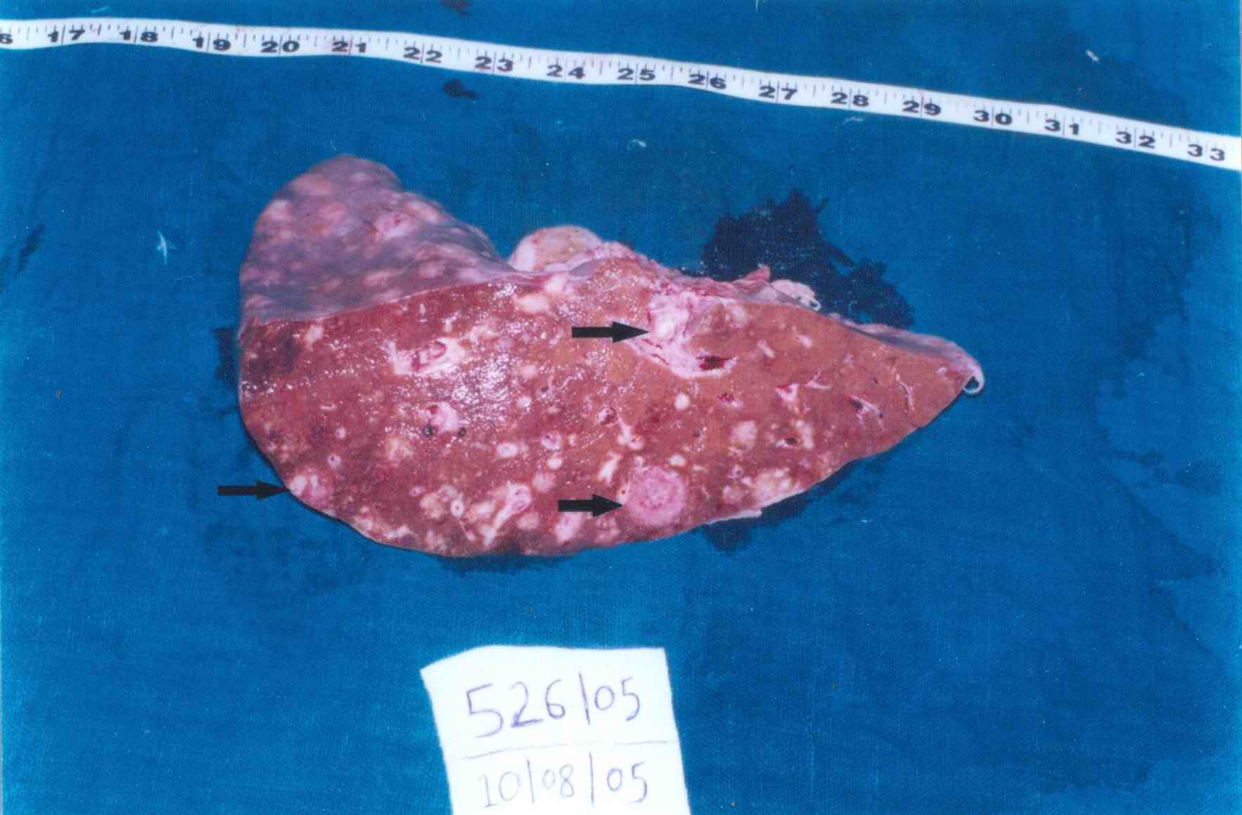
Picture 7: Liver (cut surface) shows multiple nodules of metastasis. (Please see arrows)
|
|
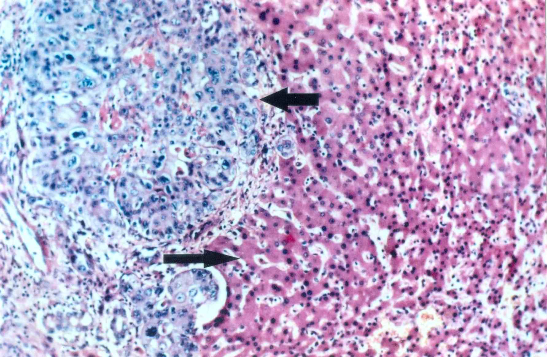
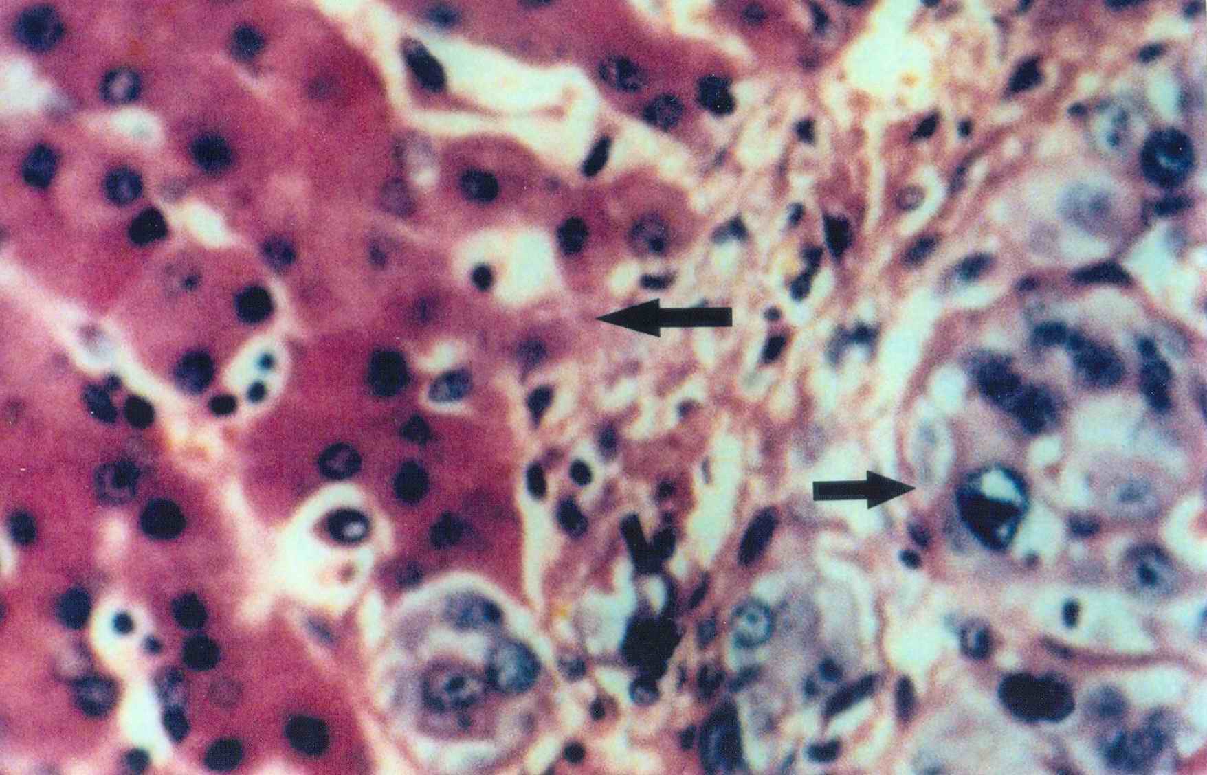
Picture 8: Liver - normal architecture on left half and tumour mass on right half. (X100) . Top arrow pointing to left shows normal tissue. Bottom arrow pointing to right shows infiltration of tumour cells.
|
|
Picture 9: Liver - normal architecture on left half and tumour mass on right half. (x400) . Top arrow pointing to left shows normal tissue. Bottom arrow pointing to right shows infiltration of tumour cells
|
|
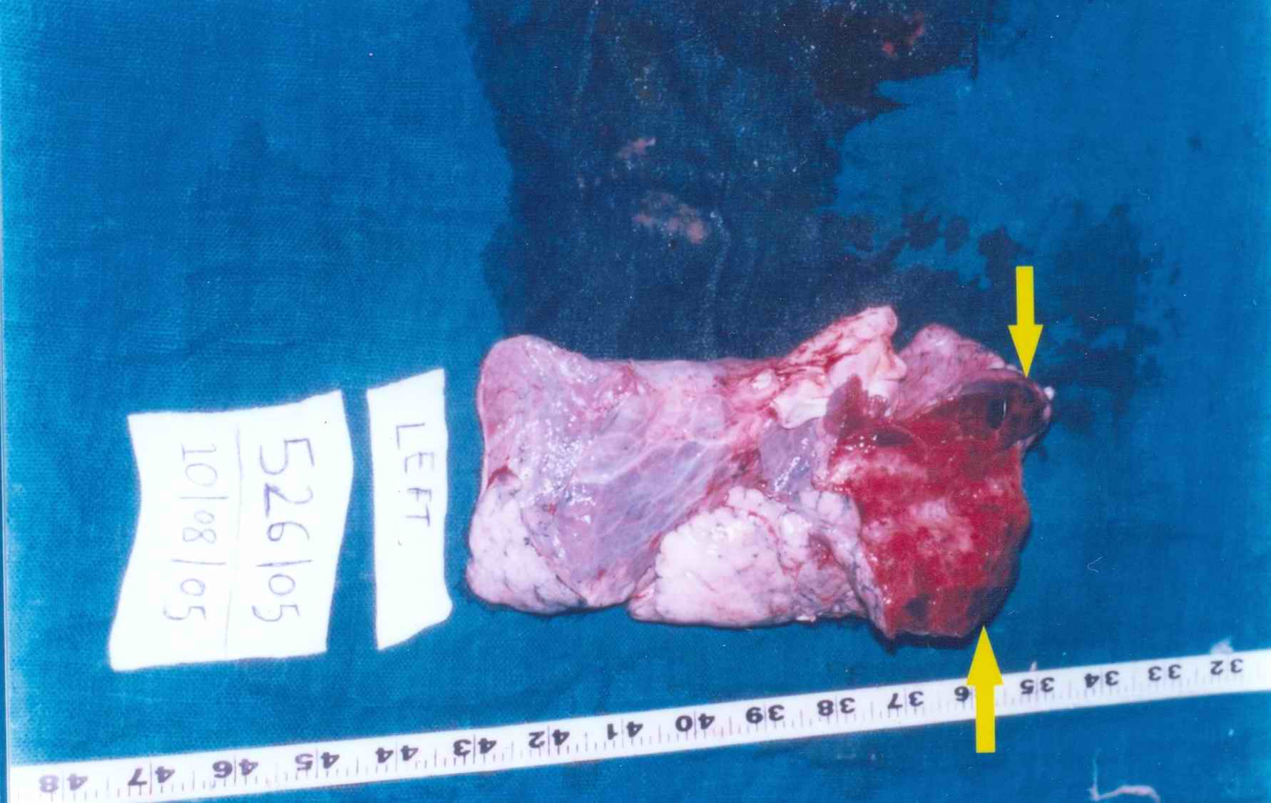
Picture 10: Lung (gross) - upper portion shows firm area, which is cut. Arrow shows nodules of metastasis.
|
|
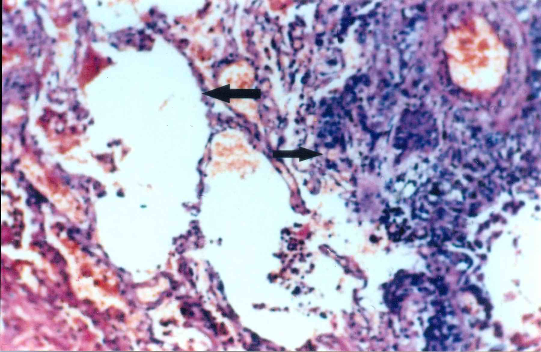
Picture 11: Lung - left half shows lung tissue and right half shows infiltration of tumour cells. (x100)
|
|
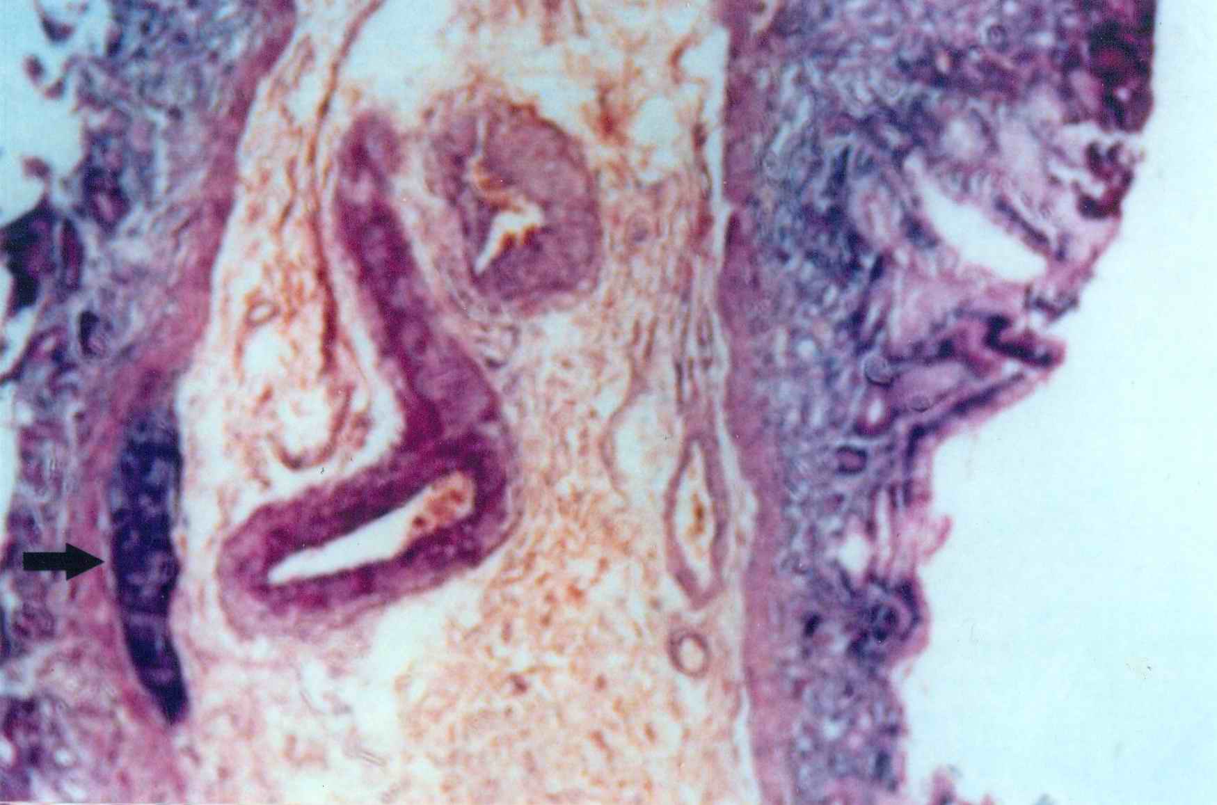
Picture 12: Stomach wall shows islands of tumour cells. (x100) (see arrow)
|
Please click all pics to enlarge
A case of carcinoma of hypopharynx was brought to the casualty department of G.G. hospital, Jamnagar, Gujarat. The patient was found dead on arrival.
A medico legal postmortem examination was conducted. Photographs are displayed to show gross and microscopic appearance of secondary metastasis in the heart and other organs. Here our main emphasis lies on the cardiac metastasis, which is a very rare event.
(N.B. Click all pictures to enlarge!)
*Corresponding author and requests for reprints:
B.D. Gupta
Professor of Forensic Medicine
M.P.Shah Medical College
Jamnagar 361008
India

Click here to contact us.
 This page has been constructed and maintained by Dr. Anil Aggrawal, Professor of Forensic Medicine, at the Maulana Azad Medical College, New Delhi-110002. You may want to give me the feedback to make this pages better. Please be kind enough to write your comments in the guestbook maintained above. These comments would help me make these pages better.
 IMPORTANT NOTE: ALL PAPERS APPEARING IN THIS ONLINE JOURNAL ARE COPYRIGHTED BY "ANIL AGGRAWAL'S INTERNET JOURNAL OF FORENSIC MEDICINE AND TOXICOLOGY" AND MAY NOT BE REPOSTED, REPRINTED OR OTHERWISE USED IN ANY MANNER WITHOUT THE WRITTEN PERMISSION OF THE WEBMASTER
Questions or
suggestions ? Please use ICQ 19727771
or email to
dr_anil@hotmail.com
Page Professor Anil Aggrawal via
ICQ
