|
|
|
 Forensic Pathology Reviews, Vol. 1, Edited by Michael Tsokos. Hard Bound, 6" x 9".
Humana Press Inc., 999 Riverview Drive, Suite 208, Totowa, New Jersey 07512; Publication Date 15 April, 2004. xii + 365 pages, ISBN 1-588-29-414-5. Price $99.50
 Official Site:Click here to visit
 |
This is one of those rare high quality books, that won critical acclaim from all four of our reviewers. The fact that they come from three different continents, confirms that this book has a global appeal. The board of editors decided to run some excerpts from this immensely useful book, so one could judge what a valuable addition to forensic literature this book has been.
 |
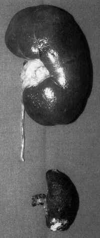
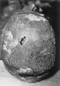
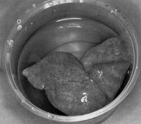
The book comprises of 15 chapters all relating to vastly different topics, covering almost the entire gamut of forensic pathology. One of the chapters that I liked most was the first entitled Morphological Findings in Burned Bodies [written by Michael Bonhert of the Institute of Forensic Medicine, University Hospital of Freiburg, Germany]. This could perhaps be because we see so many burning cases almost on a daily basis. Most of these of course come to us as cases of "dowry deaths". We do see artifactual epidural hemorrhages in several of these cases and a number of other findings described in this chapter. Yet it was a pleasure to read this chapter, if only to recapitulate what we see almost on a daily basis. Here is what Bonhert has to say on findings in Gastrointestinal tract, bones, cranial cavity and brain (pages 18-19)...
Gastrointestinal TractThe abdominal organs are protected by the abdominal wall from direct damage by the flames for a relatively long period of time. As a result of the heating of the body, the tissue fluids boil away and the pressure inside the hollow organs and the abdominal cavity builds up, which often leads to the rupture of the abdominal wall and the prolapse of intestinal loops. Consumption of the abdominal wall by the fire further promotes the rupture. In rare cases, heat-related ruptures of the gastrointestinal organs are found, before they were directly exposed to the fire. Schneider reported on the rupture of the stomach in a 2-year-old child and a rupture of the colon in a 3-year-old child. In both cases, the abdominal wall was charred without exposing the abdominal cavity. In a case from our own autopsy material, in which the victim had remained in a sauna for several hours after death, there were ruptures of the large intestine with several liters of fluid in the abdominal cavity.
Berg and Schumann described a heat hematoma of the stomach. In that case, in which the gastric wall was actually charred, a layer of blood had been found on the chyme. The swallowing of blood could be ruled out. The authors assumed that the heat hematoma could have been promoted by strong congestion of the blood vessels in the gastric wall owing to hyperemia during digestion. BonesBones are highly resistant to heat in their gross structure and usually allow macroscopic assessment even after the body was exposed to high temperatures for several hours. At temperatures above 7000C, complete combustion of the organic substances with incineration and recrystallization of the inorganic matter occurs, which is called "calcination". The bones are grayish-whitish, desiccated, and disintegrate easily. The surface shows characteristic tears, partly reflecting the course of the trabeculae but often also being irregular in structure. As all other organs, bones can also shrink in the heat. In long bones, the reduction in length can be up to 10%. Both von Hofmann and Merkel described the problems of differentiating between vital and postmortem fractures. On the one hand, fractures may be caused by the direct effect of the fire. On the other hand, they may have occurred after death, for example, when walls or timber collapsed. Especially in charred or calcined bones, a minor mechanical strain may be enough to cause fractures. Therefore, in fractures localized within charred bone areas, the possibility of artifacts should be considered. On the other hand, several authors have stressed expressly that injuries sustained during life can be demonstrated even on charred or calcined bones. Eckert et al. found a fracture of the iliac bone caused by blunt force on the pelvis of a female body largely consumed by fire. Merkel and also Henmann pointed out that traces of sharp force especially can be demonstrated quite well on the skeleton of burned bodies. Fractures away from areas of burned bone are in all probability not a result of thermal effects.
In the assessment of burned bones, special attention must be paid to the bony skull cap. In about one-third of all fire deaths, the skull cap is partially destroyed and the interior of the skull is exposed (8), which makes assessment even more difficult. Isolated fractures of the external table are seen, especially in those cases where a defined area of the skull cap was in direct contact with the flames. Prolonged exposure to heat causes fractures of the entire thickness of the skull cap with occasional bursting of the sutures of the skull. The tears in the skull cap caused by heat may radiate from a center, but can sometimes also be elliptic or circular in shape or resemble a spider's web fracture. In rare cases, round or oval bone fragments may burst outward. Distinction of this finding from a gunshot injury may be difficult. Careful examination and consideration of the other findings and the circumstances of the case allow differentiation of mechanical trauma sustained during life from postmortem trauma or heat artifacts, even when consumption by the fire is far advanced. Cranial Cavity and BrainA frequent finding is the epidural hematoma caused by heat. This is a postmortem effect resulting from the shift of fluid from the diploe and the venous sinuses when the skull cap is in direct contact with the flames. Accordingly, charring of the bony skull is usually found above the site of the heat hematoma. Less often the heat hematoma is localized on the side opposite to the site where the fire had its maximum effect. It is dry, crumbly, and of brick-red color. Occasionally it may be surrounded by fat, and in rare cases accumulations of fat without extravasations of blood can also be found in the epidural space. Apart from that, the hematoma is sometimes found to be interspersed with brain tissue when the dura mater is torn owing to shrinkage by heat. The shift of brain tissue into the epidural space is caused by the elevated steam pressure in the cranial cavity resulting in an enlargement of the brain. |
||
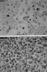
Another chapter that catches immediate attention is chapter 3 entitled Timing of Cortical Contusions in Human Brain Injury: Morphological Parameters for a Forensic Wound-Age Estimation by Roland Hausmann of the Institute of Legal Medicine, Friedrich-Alexander University, Erlangen. We all know how important it is to time the wounds for legal purposes. For cortical injuries, the problem is far from solved. This chapter attempts to summarize the knowledge attained till the present time. Here is what the author has to say at page 59:
Macrophage-Microglial reactionsIn recent years, it has been recognized that the major component of the response to nerve injury derives from microglia cells and macrophages. Cerebral macrophages were first described by Nissl and extensively evaluated by Rio-Hortega. Today it is widely accepted that this cell type is ontogenetically related to the monocytic lineage and invades the CNS during fetal development. Microglia, the resident macrophages of the CNS, take on an activated phenotype: the typical ramified microglial cells adopt an amoeboid form, thus they cannot be discriminated from infiltrating monocytes. Furthermore, there is evidence that activated microglia upregulates its expression via cell surface antigen molecules, such as the leukocyte common antigen vimentin, CD4, and the major histocompatibility (MHC) antigens class I and class II. Because the distribution as well as the number and functional state of cerebral macrophages depends on the survival period, information on the course of morphological and immunological features after tissue destruction can contribute to a forensic wound-age estimation.
Conventional histological studies revealed varying results concerning the temporal appearance of brain macrophages in cortical lesions: some authors observed cerebral macrophages as early as a few hours after the injury. On the other hand, a significant macrophage reaction has been reported 12-14 hours or 1-2 days after the injury at the earliest. A further classification of cerebral macrophages according to the different kind of incorporated material can also be useful for estimating the age of a traumatic brain lesion. Macrophages containing hemosiderin (siderophages) have been found in cortical lesions in cases with survival periods of at least 2-5 days whereas the non-iron-containing pigment hematoidin was detectable 1-2 weeks after the injury at the earliest. Macrophages showing fatty granules could be observed 3 days after the trauma. The macrophage activity in traumatically injured human brain was extensively investigated by Oehmichen et al. The authors described intracellular lipid deposits as early as 24 hours and regularly 5-6 days after the trauma. The minimal survival period was 10 days for the appearance of anisotropic lipids (cholesterol), 16 days for erythrophages, 71 hours for siderophages, 13 days for hematoidin, and 101 hours for ceroid. Regarding the time-dependent immunoreactivity of cerebral macrophages some experimental studies in animals demonstrated elevated numbers of acetylated low-density lipoprotein expressing microglia cells at the earliest 5-8 hours after TBI in rats. Two days after trauma, a positive immunoreactivity could be observed for MHC I as well as for MHC II and ED 1 and ED 2. |
|
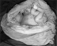
One perennial problem of forensic pathologists is to tell whether an infant was born dead, or was born alive but killed later. Tsokos has been careful to include a chapter dealing with this problem. This is chapter 6, entitled Medicolegal Problems with Neonaticide written by Roger Byard of Forensic Science Center, Adelaide, Australia. On pages 181-183 while dealing with the methods for determining live birth, this is what he has to say:
Methods For Determining Live BirthGeneral AspectsDetermination of whether an infant was born alive or dead is one of the most difficult aspects of these cases. A further problem is that the definition of what constitutes "live birth" legally differs from jurisdiction to jurisdiction. Requirements have included complete expulsion from the birth canal with a heart beat and/ or respiratory efforts. Unfortunately, an autopsy examination simply cannot determine whether a heart has functioned or whether the body was completely expelled prior to death, and so pathological opinion relies on an assessment of the degree of pulmonary inflation, the presence or absence of a vital reaction in the tissues, or evidence of feeding. The age of viability also varies among jurisdictions with 24 and 28 weeks being cited as the lower limits of potential survival.
Signs of intrauterine death, caused by a process of sterile tissue breakdown or maceration, may be present indicating that live birth has not occurred. During this process the body undergoes a series of characteristic changes beginning with reddening, slippage, and peeling of the skin after 12 hours, followed by purple discoloration and blister formation after 24 hours, and the development of pleural, peritoneal, and pericardial effusions after 48 hours. After several days the body has lost tone, joints become hypermobile, and cranial bones have collapsed producing Spalding's sign on radiography. An infant with changes of maceration has not been alive outside the uterus. The assertion that intraalveolar squames and/or meconium indicates stillbirth is not correct as these findings merely indicate that some degree of fetal distress has occurred and may be found in living infants some time after birth. The most reliable evidence of live birth is an independent and reliable witness who has either seen the infant moving or heard the infant crying. Milk within the stomach indicates that the infant was alive long enough to feed and was capable of such activity. Drying and separation of the umbilical cord stump, which occurs after 24-48 hours, with histological evidence of a tissue reaction, may also be useful, but does not help with deaths in the immediate postdelivery period. Flotation TestOne of the most time-honored tests used to assess the amount of pulmonary inflation that has occurred is the flotation test. This is based on the hypothesis that the lungs from an infant who has breathed will be expanded and filled with air and therefore will float in water, in contrast to the noninflated lungs of a stillborn infant, which will sink. Some authors suggest that it is better to attempt to float the lungs and heart en bloc to increase the sensitivity of the test.
Unfortunately, interpretation of this test is fraught with difficulty as there are numerous false positives and negatives, making this test of dubious usefulness in isolation. For example, lungs from a stillborn infant may float if there has been attempted resuscitation, with forcing of air into distal airspaces, or if there has been generation of gas within lung tissues by putrefactive bacteria. Similarly, lungs from a live-born infant may not have been inflated sufficiently to float if respiratory efforts have been weak. It has even been asserted that moving a dead infant may cause air to be aspirated into the lungs. However, though considering these caveats it can certainly be said that salmon-pink spongy lungs that float in water, in the absence of resuscitation and putrefaction, are most in keeping with lungs from an infant who has breathed. Radiographs may be used to assess the degree of pulmonary inflation and also to detect air within the stomach and upper gastrointestinal tract. If resuscitation or putrefaction have not occurred, it is assumed that air has reached the gut from swallowing. The stomach may also float in water if distended by air. The usefulness of attempting to demonstrate air within the middle ears is debatable and the relationship between the presence of pulmonary interstitial emphysema and possible live birth is yet to be clarified. Lung WeightsAnother measurement that has not proven of much use is comparison of lung to body weights. This was based on the observation that inflated and perfused lungs are heavier than lungs where respiration has not occurred. Again considerable inaccuracies occur. "Birth-Line"The so-called "birth-line" in teeth refers to a line caused by disturbance of ameloblast activity at birth that can be detected after several weeks. Although scanning electronmicroscopy has been used to identify this finding within several days of birth, its practical usefulness is not great given that most deaths occur earlier than this. |
||
Perhaps these excerpts would not be complete if I do not mention about another wonderful chapter on autoerotic deaths. Written by Stephan Seidl, it gives some remarkable photographs, besides giving an excellent table enumerating various characteristics for a determination of autoerotic death.
Here is that table. It appears on pages 256-257:
|
|||||||||||||||||||||||||||||||||||||||
In short, it is an excellent book to read. You would thank me I recommended such a beauty to you!
-Anil Aggrawal
Editor-in-Chief
 Review 1 by Erik Edston, Sweden
Review 3 by Benjamin Swift, UK
Review 4 by Ronald Wright, USA
An Exclusive interview with Michael Tsokos
Other reviews of this book:
Order Humana Press Books by clicking here.
or via telephone: (973) 256-1699 or Fax: (973) 256-8341 or Email:humana@humanapr.com
 Request a PDF file of this review by clicking here. (If your screen resolution can not be increased, or if printing this page is giving you problems like overlapping of graphics and/or tables etc, you can take a proper printout from a pdf file. You will need an Acrobat Reader though.)
N.B. It is essential to read this journal - and especially this review as it contains several tables and high resolution graphics - under a screen resolution of 1600 x 1200 dpi or more. If the resolution is less than this, you may see broken or overlapping tables/graphics, graphics overlying text or other anomalies. It is strongly advised to switch over to this resolution to read this journal - and especially this review. These pages are viewed best in Netscape Navigator 4.7 and above.
-Anil Aggrawal

 [ Major links ]
[ Aims and Objectives ] [ FAQ ] [ Editorial Board ] [ Contributing Partners ] [ Sitemap ]
[ Paper/Thesis submission guidelines ] [ Editorials - Cumulative Index ] [ Discussion ] [ Chat room ] [ Be our sponsor! ]
[ Cumulative index of Book Reviews sorted by | Publishers | General Interest Books | Technical Books ] [ Animated Reviews ] [ Featured Reviews ] [ E-books ]
[ Reviews with Quizzes ] [ Links ] [ Submit books/journals/software/multimedia for review ] [ journal CD ] [ History of the Journal ] [ Interviews ] [ Credits ]
[ Cumulative index of | Theses/dissertations | [ Online Courses ] [ Awards ] [ Anil Aggrawal's Internet Journal of Book Reviews - Sister Publication ]
[ Cumulative reviews of Software/Multimedia | Books on CD/Audio tapes ] | Calenders | Models ] [ contact us ]
[ Undergraduate section | Postgraduate section ] [ Forensic gadgets/toys/other tidbits ]
Books for review must be submitted at the following address.
Professor Anil Aggrawal (Editor-in-Chief)
Anil Aggrawal's Internet Journal of Forensic Medicine and Toxicology
S-299 Greater Kailash-1
New Delhi-110048
India
Click here to contact us.
This page has been constructed and maintained by Dr. Anil Aggrawal, Professor of Forensic Medicine, at the Maulana Azad Medical College, New Delhi-110002. You may want to give me the feedback to make this pages better. Please be kind enough to write your comments in the guestbook maintained above. These comments would help me make these pages better.
 IMPORTANT NOTE: ALL PAPERS APPEARING IN THIS ONLINE JOURNAL ARE COPYRIGHTED BY "ANIL AGGRAWAL'S INTERNET JOURNAL OF FORENSIC MEDICINE AND TOXICOLOGY" AND MAY NOT BE REPOSTED, REPRINTED OR OTHERWISE USED IN ANY MANNER WITHOUT THE WRITTEN PERMISSION OF THE WEBMASTER
Questions or suggestions ? Please use ICQ 19727771 or email to dr_anil@hotmail.com
Page Professor Anil Aggrawal via ICQ

|