|

|
|
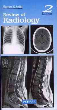
 Review of Radiology, 2nd Edition by Sumer K Sethi, Softcover, 4.5" x 8.5".
Peepee Publishers & Distributors (P) Ltd., 7/31, First Floor, Ansari Road, Daryaganj, New Delhi - 110002, India. Ph: 9811156083, 011-55195868. E-mail:peepee160@yahoo.co.in: Publication Date 2005. 201 pages, ISBN 81-88867-29-2. Price: $6.00, £3.00, Indian Rs 100.00
 Official site: http://www.peepeepub.com/default.asp
 |
This is one of those rare high quality books, that won critical acclaim from all eight of our reviewers. The fact that they come from three different continents, confirms that this book has a global appeal. The board of editors decided to run some excerpts from this immensely useful book, so one could judge what a valuable addition to forensic literature this book has been.
The book comprises of several chapters all relating to vastly different topics, covering almost the entire gamut of radiology. One of the chapters that I liked most was the first which deals with historical aspects. Here is what Sethi has to say on page 1...
DISCOVERY. X-Rays were discovered by: W.C. ROENTGEN (GERMANY) In the latter half of the year 1895, a German scientist called Roentgen was working in his laboratory at the Physical Institute of the University of Wurzburg, Germany, experimenting with a type of discharge tube called Crooke's tube. The tube displayed a fluorescent glow when a high voltage current was passed through it. When he shielded the tube with heavy black cardboard, he found that a greenish fluorescent light could be seen on a fluorescent screen kept some 9 feet away. Roentgen concluded that a new type of ray was emitted from the tube that could pass through the black covering. The rays could pass through most substances, including the soft tissues of the body, but left the bones and most metals visible. One of his earliest photographic plates from his experiments was that of a film of his wife, Bertha's hand with a ring. Roentgen named the invisible radiations as X-rays (or unknown rays). . Discovered on Nov 8, 1895 . Roentgen got the Nobel Prize in-1901 |
This is what Sethi has to write on Pulmonary Infections on Pages 21-23 (2nd Edition)
PULMONARY INFECTIONSRadiology of Pulmonary Tuberculosis :*** Primary TB: . Ghon's lesion: Subpleural consolidation + lymphatic + enlarged lymph nodes . Lymphadenopathy is characteristic of primary infection ( also in Tuberculosis with AIDS) . Consolidation can occur anywhere in the lung. (More common subpleural sites in lower lobe) Secondary TB . Cavitation . Fibrosis . Involves Apical segments of upper and lower lobes . V. UN COMMON IN ANTERIOR SEGMENT OF UPPER LOBE** Hematogenous spread of TB leads to miliary shadowing Endobronchial spread : Tree in bud appearance** Rasmussen aneurysm: Pulmonary artery in cavity TB may cause hemoptysis** -In hemoptysis-First vessel to be studied-Bronchial artery. class="sample style2">PNEUMONIA Pneumococcal- more commonly basal, klebsiella more common right upper lobe, bulging fissure, mycoplasma earliest CXR change is fine reticulo-nodular shadows followed by consolidation. Viral pneumonias may show-peribronchial shadowing, reticulonodular shadows and consolidation Bulging Fissure . Klebsiella pneumonia (Freidlander's bacillus) . Lung abscess . Ca bronchus Hydatid Lung . No or rare calcification in lung . 'Water lily' sign OR CAMALOTE SIGN** Hydatid cyst forms three layers: Pericyst due to fibrous host reaction, ectocyst and the endocyst containing brood capsules. ASPERGILLOSIS IN LUNG • ASPERGILLOMA CXR show density surrounded by air in the cavity (air crescent sign) Also seen in - AIR Crescent sign . Aspergilloma or fungal ball . Inspissated pus in a cavity . Tumour or clot within the cavity . Hydatid cyst. • Invasive aspergillosis In immunocompromised persons CT Halo appearance due to surrounding haemorrhagic inflammation. 3) Allergic bronchopulmonary aspergillosis type III immune reaction, central bronchiectasis, 'gloved finger appearance' Pneumocystitis Carnii CXR normal in 10% may show perihilar and mid/lower zone ground glass infiltrates, lymphadenopathy pleural effusion less common. Pneumothorax is well recognized complication. Bronciectasis Morphological Types(increasing severity) 1. Cylindrical 2. Varicose 3. Cystic Radiological Signs ** • 'Signet-ring' sign- bronchus is larger than the accompanying vessel • Tram track sign- Lack of peripheral tapering (cylindrical bronchiectasis) • String of beads appearance alternate dilatation and constriction (varicoid bronchiectasis) • Cluster of grapes appearance (cystic bronchiectasis )
|
And finally sample the following lists of radiosensitivities of different tumors. Sethi gives this list on page 96 (2nd Edition). Students would find such helpful lists throughout the book.
RADIOSENSITIVITY OF DIFFERENT TUMOURSHighly sensitive
Moderately sensitive
Relatively resistant
Highly resistant
|
 Order this Book by clicking here.
Interview with Sumer Sethi.
 Request a PDF file of this review by clicking here. (If your screen resolution can not be increased, or if printing this page is giving you problems like overlapping of graphics and/or tables etc, you can take a proper printout from a pdf file. You will need an Acrobat Reader though.)
N.B. It is essential to read this journal - and especially this review as it contains several tables and high resolution graphics - under a screen resolution of 1600 x 1200 dpi or more. If the resolution is less than this, you may see broken or overlapping tables/graphics, graphics overlying text or other anomalies. It is strongly advised to switch over to this resolution to read this journal - and especially this review. These pages are viewed best in Netscape Navigator 4.7 and above.
-Editor-in-Chief


 [ Major links ]
[ Reviews with Quizzes ] [ Journal CD ] [Interviews] [ Editorials - Cumulative Index ]
[ Cumulative index of Book Reviews sorted by | Publishers | Subjects | Multimedia Reviews (cumulative) ]
[ Links ] [ Submit books/journals/software/multimedia for review ] [ Reviewers' Panel ] [ Featured Reviews ] [ Audio books ] [ My Books ]
[ Anil Aggrawal's Internet Journal of Forensic Medicine and Toxicology - Sister Publication ] [ contact us ]
Books for review must be submitted at the following address.
Professor Anil Aggrawal (Editor-in-Chief)
Anil Aggrawal's Internet Journal of Book Reviews
S-299 Greater Kailash-1
New Delhi-110048
India
Click here to contact us.
This page has been constructed and maintained by Dr. Anil Aggrawal, Professor of Forensic Medicine, at the Maulana Azad Medical College, New Delhi-110002. You may want to give me the feedback to make this pages better. Please be kind enough to write your comments in the guestbook maintained above. These comments would help me make these pages better.
 IMPORTANT NOTE: ALL REVIEWS APPEARING IN THIS ONLINE JOURNAL ARE COPYRIGHTED BY "ANIL AGGRAWAL'S INTERNET JOURNAL OF BOOK REVIEWS" AND MAY NOT BE REPOSTED, REPRINTED OR OTHERWISE USED IN ANY MANNER WITHOUT THE WRITTEN PERMISSION OF THE WEBMASTER

|