

|
 OTHER REVIEWS IN THIS ISSUE OTHER REVIEWS IN THIS ISSUE Pages: |1|
2|
3|
4|
5|
6|
7|
8|
9|
10|
11|
12|
13|
14|
15|
16|
17|
18|
19|
20|
21|
22|
23| Pages: |1|
2|
3|
4|
5|
6|
7|
8|
9|
10|
11|
12|
13|
14|
15|
16|
17|
18|
19|
20|
21|
22|
23|
[Popular Books Section]
[Animated Reviews]
|

|
TOUR DE FORCE |
 Encephalo-Peripheral Nervous System: Vascularisation - Anatomy - Imaging by André Leblanc
Springer-Verlag, GmbH & Co.KG, Tiergartenstrasse 17, D-69121 Heidelberg, Germany; 923 figures, xi + 438 pages, 2540 g: ISBN 3-540-67843-3. Publication Date 2001: Hardcover Price: DM 597.06, sFr 513.60, £ 206.00, $ 299.00
 |
|
The study of anatomy by charts and diagrams is not a new technique. It has been tried time and over again, but the trick is to produce a book with clear and accurate diagrams. This is not as easy as it sounds. Human anatomy is so complex that to depict each and every structure in great detail is quite difficult, if not impossible. One of the most successful persons in this respect has been the tireless Frank H. Netter, who produced a series of highly accurate anatomical diagrams for Ciba Collection of Medical Illustrations. Needless to say such a work can best be done by a person who is both a trained artist and an anatomist. This combination is not easy to find. But Netter was one such person. The other person of course is André Leblanc, who has produced the book under review.
 |
I had heard about André Leblanc, and his book, but had never seen it. When this book arrived for review on a Saturday afternoon, initially I found myself thumbing it lazily, but very soon arose from my slumber. The high quality of the book, its production, its diagrams was striking. It is in fact one of the best anatomy books that I have seen. And it is much more than what has been produced in this genre before. For it gives not just diagrams but even photographs from actual anatomical specimens, radiographs, CT scan pictures, MRI pictures and much more.
 |
Frequently the same anatomical area is shown from two or more different viewpoints. On page 178 for instance (see figure on the left), you see not just an anatomic section at the level of mandibular nerve showing inferior alveolar nerve until mandibular canal, but also how the same area would look in an MRI image. Very few anatomy atlases have treated anatomy from this multidisciplinary approach. What you see in actual anatomical dissections is sometimes quite different from what you see in an MRI or CT image, and it is not always easy to correlate the two. By providing such comparison pictures, this book makes the task of radiologists very easy and is thus an excellent book for radiologists, neurologists and general clinicians.
 |
Sometimes the author gives not just two, but more different viewpoints for the same anatomical area. For instance on page 10 (see figure on the right), he is trying to show olfactory pathways. He shows these pathways not just in a diagram, but actually superimposes the diagram over a CT scan! And he doesn't stop at just that. He goes on to give a picture of a real anatomical specimen of the same area and also an MRI image of that area. This amalgamation of several disciplines is what makes this book truly unique.
 |
At other places dissected anatomical specimens have been juxtaposed with crisp diagrams making the understanding of various structures very easy. Take for example Pterygopalatine ganglion and fossa. As a medical teacher I do realize that this is one of the most confusing structures for students to comprehend and dissect. But the way this atlas shows this structure both in a real anatomical specimen and a diagram makes is for everybody very easy to comprehend. This comparison diagram appears on page 166 (see figure on the left). Such pictures make it a very good book for students, for not only can they understand anatomy better, but they would know how to draw the relevant diagrams in their exams to get good grades.
Does this book deal with the whole of human anatomy? No, it doesn't, but I wish one day Leblanc would think of producing such a book too. The current book however deals with just the twelve cranial nerves. Whole of their course right from their real origin from the nuclei within the brain through their intracanalicular and intracranial course to their ultimate destination - various muscles, organs or other structures - is traced. Anatomic variations such as asymmetry and vulnerable points are considered. Bony orifices occupied by cranial nerves are each investigated precisely along their axis (taking possible asymmetry into account) thanks to the reference and centering angles devised by the author himself.
 |
 |
How detailed the treatment of each nerve is can be gauged by the fact that just these 12 cranial nerves have been dealt with in 923 figures - an astounding average of 77 diagrams per cranial nerve! The book spans 438 pages, again devoting an average of about 36 pages per nerve.
Each nerve is dealt with under a number of different headings. Take for example the Vagus nerve. It is dealt with under the following sections: 1. Anatomy (Course - Distribution - Collaterals) 2. Real origin of Vagus nerve 3. Apparent origin of vagus nerve 4. Jugular foramen 5. Ostium Introitus and sulcus of Auricular branch 6. Courses of Vagus nerves (both) and 7. Arteries for vagus nerve.
The good news for those who flinch at the idea of reading thick tomes is that the book doesn't offer much for reading. The writer seems to believe in the old Chinese saying," A picture says more than a thousand words". The whole of cranial nerve anatomy is explained by way of pictures of actual anatomical specimens, CT and MRI images, diagrams, charts and a host of other different views such as subtraction radiography and so on.
 |
 |
At the beginning of each chapter, a big diagram of the relevant nerve (drawn by Leblanc himself) appears, which sets the stage - so to say - for an exploration of that nerve. I found myself peering at these interesting diagrams for several minutes, and honestly was quite conversant with the nerve just by seeing this diagram. This does make the subsequent exploration quite painless. For the benefit of readers, I am reproducing just two such diagrams here - of the fifth nerve appearing on page 79 and of seventh nerve appearing on page 227.
Some anatomical sections are so well prepared that they are sheer pleasure to look at. Take for instance the anatomical preparation of the brain stem prepared by Prof. J.P.Francke, of the faculty of Medicine, Lille, which appears on page 232. It shows the apparent origin of each and every cranial nerve so clearly that it is difficult to forget it afterwards. In fact this is the diagram which I found myself returning to again and again to refer to any nerve.
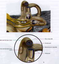
Another good feature of the book is the several fenestration diagrams, the author has produced himself. For instance, on page 356, he "cuts" the semicircular canals at various points and shows how the membranous labyrinth would look from those "little windows". At other places we see arteries of trigeminal nerve through fenestrations showing trigeminal ganglion. I found diagrams like these particularly useful, as they allow you to see structures in an entirely new perspective and help understand the anatomy of this region quite effortlessly.
 |
Some interesting trivia also appear at several places in specially made boxes. Which is the thinnest cranial nerve? And which is the thickest? Trochlear and Trigeminal respectively. Such pieces of information make the text very lively and interesting.
Leblanc has also produced a number of illustrative posters relating to cranial nerves. Eight such posters appear at the end of this book, and I am reproducing just one here, so readers can have an idea what I am talking about. The actual size of these posters is 69 x 85 cm, and I am told these charts are displayed in several radiology and other departments all over the world. Readers who might be interested in these charts can contact the author directly (Mr. André Leblanc, P.O. Box no. 2, 80800 Daours, France. Fax: +33 3 22.48.31.23)
 |
Whom is the book meant for? Well, I would imagine, anyone who is connected with the study of human body in any way would be interested in this book. But the professionals who would be most interested in this book would be neurologists, neurosurgeons, neuroradiologists and neuroanatomists, for this book deals with cranial nerves. It would be useful for general pathologists and forensic pathologists like me, who have to frequently deal with cerebral injuries and identify the injured cranial nerves. These do have severe medicolegal repercussions. Maxillofacial surgeons, odontostomatologists, otolaryngologists, ophthalmologists and dentists would find it useful for most of their concerned "areas" is covered in this book. This would definitely be one of my most valuable possessions, and I would frequently be referring to it when dealing with cranio-cerebral injuries.
 Order Encephalo-Peripheral Nervous System: Vascularisation - Anatomy - Imaging by Clicking here
 Request a PDF file of this review by clicking here. (If your screen resolution can not be increased, or if printing this page is giving you problems like overlapping of graphics and/or tables etc, you can take a proper printout from a pdf file. You will need an Acrobat Reader though.)
N.B. It is essential to read this journal - and especially this review as it contains several tables and high resolution graphics - under a screen resolution of 1600 x 1200 dpi or more. If the resolution is less than this, you may see broken or overlapping tables/graphics, graphics overlying text or other anomalies. It is strongly advised to switch over to this resolution to read this journal - and especially this review. These pages are viewed best in Netscape Navigator 4.7 and above.
-Anil Aggrawal

 [ Major links ]
[ Aims and Objectives ] [ FAQ ] [ Editorial Board ] [ Contributing Partners ] [ Sitemap ]
[ Paper/Thesis submission guidelines ] [ Editorials - Cumulative Index ] [ Discussion ] [ Chat room ] [ Be our sponsor! ]
[ Cumulative index of Book Reviews sorted by | Publishers | General Interest Books | Technical Books ] [ Animated Reviews ] [ Featured Reviews ] [ E-books ]
[ Reviews with Quizzes ] [ Links ] [ Submit books/journals/software/multimedia for review ] [ journal CD ] [ History of the Journal ] [ Interviews ] [ Credits ]
[ Cumulative index of | Theses/dissertations | [ Online Courses ] [ Awards ] [ Anil Aggrawal's Internet Journal of Book Reviews - Sister Publication ]
[ Cumulative reviews of Software/Multimedia | Books on CD/Audio tapes ] | Calenders | Models ] [ contact us ]
[ Undergraduate section | Postgraduate section ] [ Forensic gadgets/toys/other tidbits ]
Books for review must be submitted at the following address.
Professor Anil Aggrawal (Editor-in-Chief)
Anil Aggrawal's Internet Journal of Forensic Medicine and Toxicology
S-299 Greater Kailash-1
New Delhi-110048
India
Click here to contact us.
This page has been constructed and maintained by Dr. Anil Aggrawal, Professor of Forensic Medicine, at the Maulana Azad Medical College, New Delhi-110002. You may want to give me the feedback to make this pages better. Please be kind enough to write your comments in the guestbook maintained above. These comments would help me make these pages better.
 IMPORTANT NOTE: ALL PAPERS APPEARING IN THIS ONLINE JOURNAL ARE COPYRIGHTED BY "ANIL AGGRAWAL'S INTERNET JOURNAL OF FORENSIC MEDICINE AND TOXICOLOGY" AND MAY NOT BE REPOSTED, REPRINTED OR OTHERWISE USED IN ANY MANNER WITHOUT THE WRITTEN PERMISSION OF THE WEBMASTER
Questions or suggestions ? Please use ICQ 19727771 or email to dr_anil@hotmail.com
Page Professor Anil Aggrawal via ICQ

|
