

|
 OTHER REVIEWS IN THIS ISSUE OTHER REVIEWS IN THIS ISSUE Pages: |1|
2|
3|
4|
5|
6|
7|
8|
9|
10|
11|
12|
13|
14|
15|
16|
17|
18|
19|
20|
21|
22|
23| Pages: |1|
2|
3|
4|
5|
6|
7|
8|
9|
10|
11|
12|
13|
14|
15|
16|
17|
18|
19|
20|
21|
22|
23|
[Popular Books Section]
[Animated Reviews]
|

|
AN INTERACTIVE BOOK ON ANATOMY |
 Kinetic Anatomy, 1st Edition by Robert S. Behnke, Paperback, 8.5" x 11"
Human Kinetics, P.O. Box 5076, Champaign, Illinois 61825-5076; 296 pages: ISBN 0-7360-0016-X. Publication Date February 2001: Price, $34.00.
 |
Tennis elbow, Golfer's elbow, Boxer's fracture, baseball finger, skier's thumb.. .. .. Name a sport, and there is an eponymous injury associated with it. What are these injuries? Which structures are affected? Why are they caused in the first place? How can they be prevented? And once you get it, what can be done to relieve them? These are the questions which concern a sport physiotherapist today. So much is sport medicine relevant today that every sports team worth its salt has its own physician, physiotherapist, psychologist and other medical and paramedical personnel dedicated and committed to problems associated with that particular sport.
How best can these medical and paramedical personnel gain knowledge of human anatomy, which is relevant to them? Or how does a student of such disciplines as kinesiology, biomechanics, physiology, exercise physiology, neuroanatomy and rehabilitation techniques learn anatomy in a most painless way? The book under review comes as a succor to all these students and practitioners. It attempts to give anatomy which is relevant to sports professionals, and it gives it in a very interesting, painless, interactive manner.
The book is designed as an entry-level text for those who have little or no background or academic experience in human anatomy. The book has threefold aims, as the author sets out very clearly in the preface of the book. The first goal is to present the basic vocabulary of anatomy to the beginner. This knowledge enables readers to communicate with colleagues, physicians and therapists. To this end, the author gives a box called "Review of Terminology" at the end of each chapter. This box enumerates all special anatomical terms that have made an appearance in that chapter.
|
The second aim of the book is to give readers a firm concept of how a human body is constructed and how it moves by discussing various structures involved in movement, such as bones, joints, and muscles. Knowing what structures are involved and how they should be functioning allows an individual to identify problems and correct them to enhance physical activity.
The third aim is to impart knowledge that allows the pursuit of healthy living. The author confesses here that this aim is less academic, but perhaps most important of all. I would tend to agree with the author completely on this point. At one point, the author summarizes his philosophy in these words, "Human anatomy is a fascinating subject if for no other reason that that it is about you." This coincides completely with my own philosophy. I am a forensic pathologist basically, but when I dissect a human body even for medico-legal purposes, I tend to dissect each organ in somewhat greater detail than my work would normally demand. Often my students ask me why I do this, and I am usually unable to give a satisfactory answer to them. Well, I guess, I am fascinated by human anatomy because it is about me, and when I am dissecting a cadaver, I am actually trying to discover, what lies inside my own body.
The book has thirteen chapters divided into four parts. Part I, having two chapters discusses general concepts of anatomy. Part II runs from chapter 3 to 6 and discusses the upper limb. Part III discusses the spinal column, pelvis and thorax and spans three chapters. Remaining four chapters are in part IV, dealing with lower extremity. Thus almost the whole of anatomy is dealt with. At the end of each part there is an extensive summary table that includes muscle origins, insertions and actions and nerve and blood supply. These tables provide a ready reference for the material covered in the chapters of each part.
One feature of the book that interested me most is a section called "Suggested Learning Activities", which appears at the end of each chapter. These interactive activities help to consolidate various anatomical concepts in the mind of the student. Take for example this learning activity which appears on page 59, at the end of chapter 3 (The Shoulder).
Place your hands on a partner's scapula. Ask the partner to slowly abduct both shoulder joints. As the humerus moves away from the body, determine when the scapula starts to move. Did the scapula move throughout abduction of the shoulder joint? When did it start to move? Why did it move? What muscle initiated this action?
Or sample this learning activity, which appears on page 220, at the end of chapter 11 (The Knee).
Sitting up on your table with your lower legs off the end of the table and your feet next to each other, extend both knee joints to full extension. As your knees moved to full extension, what did the lower legs (and the relationship of your feet to each other) do?
I attempted several of these activities, and before I realized it, I had learnt several important concepts of anatomy. And all along I didn't know I was learning; I thought I was having fun!
Another interesting feature is the Multiple Choice Questions (MCQs), which appear at the end of each chapter. This approach tests the retaining power of the students. Another additional exercise that appear at the end of each chapter is "Fill-in-the-Blank questions". Trying out these MCQs and "Fill-in-the Blanks" helps students to judge for themselves how much they have retained. But as the author warns in his preface, "you should take this practice exam only after you have studied the material".
|
The book is interspersed with several interesting and illustrative sidebars. One sidebar that I liked most was a special box called "Focus on.. ." This box appears off and on throughout the book, and gives important practical points related to the structure the author is discussing at that particular place. Not only does it help the student to relate the anatomy with important practical aspects, but it also gives important tips on how best to avoid that injury. Sample this box which appears on page 149.
Focus on.. ..
The crest of the ilium is the source of many muscle attachments, both origins and insertions. When a person contuses this area, it is very painful to attempt to move the lower spinal column and the hips and even to attempt to breathe heavily because of the muscular attachments. This type of trauma to the soft tissue attaching to the crest of the ilium is often referred to as a "hip pointer."
Injuries such as a hip pointer can sometimes be the result of improperly fitted or non-use of equipment. All coaches should have instructions in which the proper use and proper fitting of equipment are discussed. Knowledge of specific anatomical structures and how equipment protects those structures can reduce the incidence and severity of some injuries.
The Crest of the Ilium
The following box appears on page 237
Focus on.. ..
Spraining the ligaments of the first (great toe) MP (MetatarsoPhalangeal) joint (often from hyperextension) is frequently referred to as "turf toe," depending on the mechanism of injury. Study of footwear and its interaction with playing surfaces can help with understanding how to prevent this minor but debilitating condition. Knowledge of anatomy, physics, and biomechanics, combined with the experience of engineers familiar with the effects of the interaction of footwear and the playing surface, can assist in preventing the sprained first MP joint.
Turf Toe
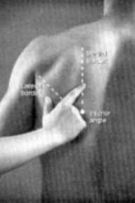 |
Another very useful feature is that the author tries to show various structures of the body in an actual body. This helps the students to understand better, where a particular structure is to be located. This approach is not adhered to by many anatomy books. They are content to show the anatomical structures in numerous line diagrams, or otherwise. The result is that the student often crams up those diagrams, and although able to reproduce those diagrams in an examination, is completely ignorant of that structure in an actual body. Sample this diagram on the left, which appears on page 38 of the book. In this photograph of the back of an actual person, the author tries to show the lateral border, medial border and inferior angle of the scapula. In normal anatomy books, the same borders and angles would be shown on the diagram of a bone. The student would normally cram up these facts, but would remain completely ignorant of the actual location of these structures in a human body.
 |
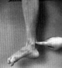 |
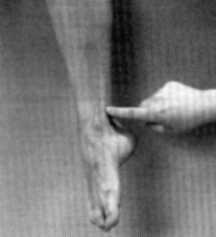
Or sample these two diagrams (on the right) which appears on page 244. In the first diagram, the author shows the location of tibialis posterior muscle, while in the second diagram, the position of flexor digitorum longus is shown. Such photographs which constantly appear throughout the book, are very good learning aids.
This "Hands-on" approach was first followed by Joseph E. Donnelly in his book Living Anatomy, of which the present book is a descendant. This approach encourages the readers to use one-another as live models to learn anatomy.
I would fully recommend this book to all students of anatomy and physiology. The book of course is primarily meant for sport professionals and for students of kinesiology, biomechanics, physiology, exercise physiology, neuroanatomy and rehabilitation techniques. The text however is so illustrative and useful, that I would recommend it to students of general medicine, pathology and forensic pathology too. I would have loved to read this book, when I was learning anatomy in my medical school, way back in 1973-74, at the University College of Medical Sciences, New Delhi. I read Gray's Anatomy instead, which undoubtedly is the Bible of Anatomy, but tends to get monotonous, impersonal and too severely academic at times for the comfort of an average student. On the contrary, the book under review is not as detailed as Gray's, but gives practical aspects of anatomy in a no-nonsense manner and most importantly in a fun-filled way.
Still worried about those terms bandied about at the beginning of this review? Well, tennis elbow is the common man's name for Lateral epicondylitis, and is seen most often in tennis, although some other sports are also not exempt from it. It can also be seen in wheelchair athletes, especially marathon runners, as they have to repetitively pronate and flex their wrists as the elbow extends during the "push phase" of their wheelchair. Other sports in which this injury is seen are gold, baseball, swimming and a number of throwing field events such as discuss and javelin. The common denominator in all these sports is a repetitive wrist motion and a heavy torque. The muscle most frequently affected is extensor carpi radialis brevis. Signs and symptoms include pain over the anterior aspect of lateral epicondyle. Majority of tenderness is located at the origin of extensor carpi radialis brevis tendon.
Golfer's elbow? This is more often seen in golfers, although the condition is also seen in some other sports. This condition is also associated with repetitive wrist flexor and pronator muscle activity. Activities most likely to cause this condition are golf swings, baseball pitchings, overhead tennis serves and forehand racket motions. Sign and symptoms include pain and mild swelling over the medial epicondyle.
What about Boxer's fracture, baseball finger and skier's thumb? Should I go on.. .. or may be you would like to find out yourself from the book? I am sure you would go for the second option.
-Anil Aggrawal
[ Paper/Thesis submission guidelines ] [ Editorials - Cumulative Index ] [ Be our sponsor! ]
[ Cumulative index of Book Reviews sorted by | Publishers |
General Interest Books |
Technical Books ] [ Animated Reviews ] [ Featured Reviews ]
[ Links ] [ Submit books/journals/software/multimedia for review ] [ journal CD ] [ History of the Journal ] [ Interviews ] [ Credits ]
[ Online Courses ] [ Awards ] [ Anil Aggrawal's Internet Journal of Book Reviews - Sister Publication ]
[ Cumulative reviews of Software/Multimedia | Books on CD/Audio tapes ] [ contact us ]
 Order Human Kinetic Books by clicking here.
 Request a PDF file of this review by clicking here. (If your screen resolution can not be increased, or if printing this page is giving you problems like overlapping of graphics and/or tables etc, you can take a proper printout from a pdf file. You will need an Acrobat Reader though.)
N.B. It is essential to read this journal - and especially this review as it contains several tables and high resolution graphics - under a screen resolution of 1600 x 1200 dpi or more. If the resolution is less than this, you may see broken or overlapping tables/graphics, graphics overlying text or other anomalies. It is strongly advised to switch over to this resolution to read this journal - and especially this review. These pages are viewed best in Netscape Navigator 4.7 and above.


[ Major links ]
[ Aims and Objectives ] [ FAQ ] [ Editorial Board ] [ Contributing Partners ] [ Sitemap ]
Books for review must be submitted at the following address.
Professor Anil Aggrawal (Editor-in-Chief)
Anil Aggrawal's Internet Journal of Forensic Medicine and Toxicology
S-299 Greater Kailash-1
New Delhi-110048
India
Click here to contact us.
This page has been constructed and maintained by Dr. Anil Aggrawal, Professor of Forensic Medicine, at the Maulana Azad Medical College, New Delhi-110002. You may want to give me the feedback to make this pages better. Please be kind enough to write your comments in the guestbook maintained above. These comments would help me make these pages better.
 IMPORTANT NOTE: ALL PAPERS APPEARING IN THIS ONLINE JOURNAL ARE COPYRIGHTED BY "ANIL AGGRAWAL'S INTERNET JOURNAL OF FORENSIC MEDICINE AND TOXICOLOGY" AND MAY NOT BE REPOSTED, REPRINTED OR OTHERWISE USED IN ANY MANNER WITHOUT THE WRITTEN PERMISSION OF THE WEBMASTER

home
> Volume 2, Number 2, July - December 2001
> Reviews
> Technical Books > page 22: Kinetic Anatomy (you are here)
Navigation ribbon